. 2014 Oct;32(10):2690-701.
doi: 10.1002/stem.1760.

Activating receptor NKG2D targets RAE-1-expressing allogeneic neural precursor cells in a viral model of multiple sclerosis

Affiliations

Activating receptor NKG2D targets RAE-1-expressing allogeneic neural precursor cells in a viral model of multiple sclerosis

Jason G Weinger et al. Stem Cells. 2014 Oct.

Abstract

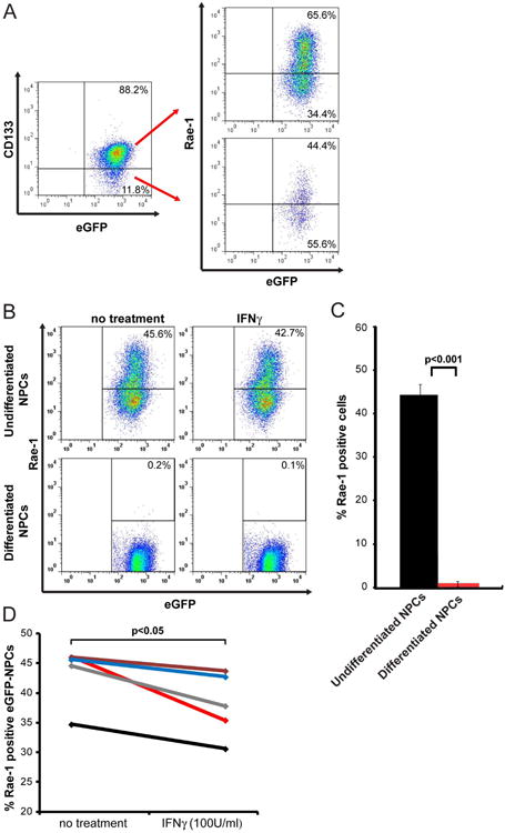
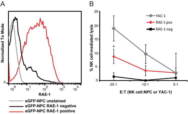
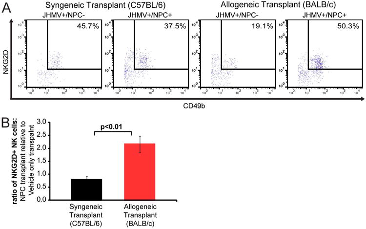
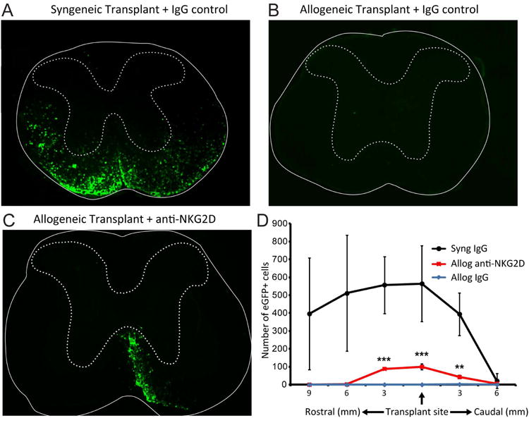
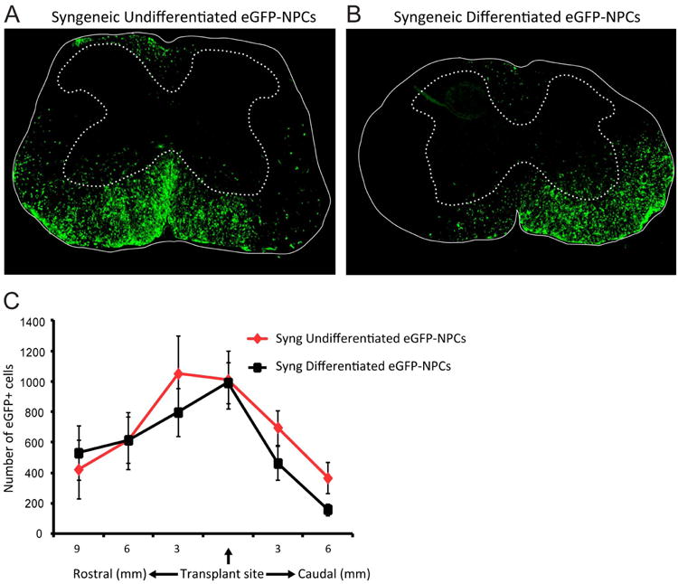
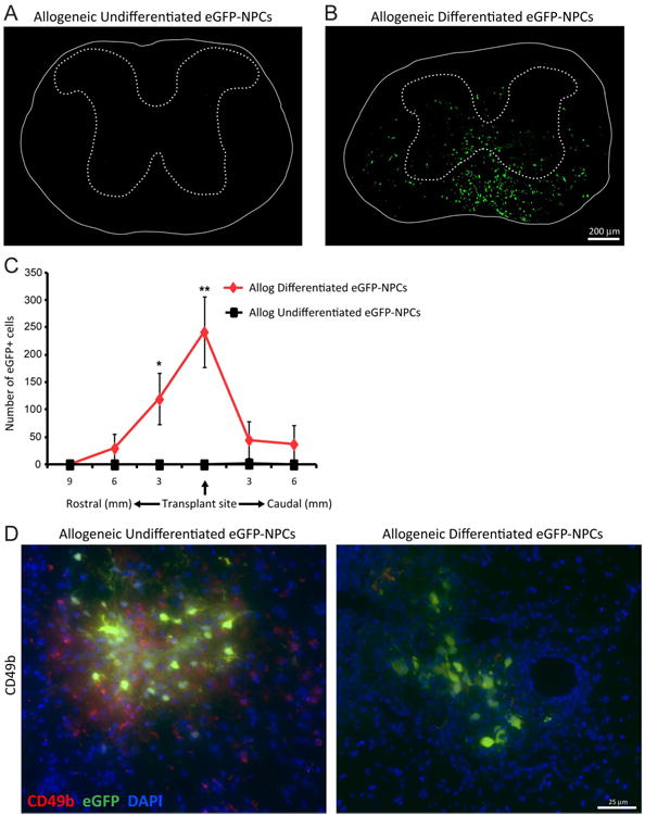
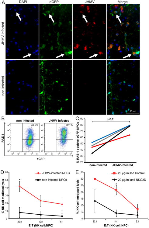
Transplantation of major histocompatibility complex-mismatched mouse neural precursor cells (NPCs) into mice persistently infected with the neurotropic JHM strain of mouse hepatitis virus (JHMV) results in rapid rejection that is mediated, in part, by T cells. However, the contribution of the innate immune response to allograft rejection in a model of viral-induced neurological disease has not been well defined. Herein, we demonstrate that the natural killer (NK) cell-expressing-activating receptor NKG2D participates in transplanted allogeneic NPC rejection in mice persistently infected with JHMV. Cultured NPCs derived from C57BL/6 (H-2(b) ) mice express the NKG2D ligand retinoic acid early precursor transcript (RAE)-1 but expression was dramatically reduced upon differentiation into either glia or neurons. RAE-1(+) NPCs were susceptible to NK cell-mediated killing whereas RAE-1(-) cells were resistant to lysis. Transplantation of C57BL/6-derived NPCs into JHMV-infected BALB/c (H-2(d) ) mice resulted in infiltration of NKG2D(+) CD49b(+) NK cells and treatment with blocking antibody specific for NKG2D increased survival of allogeneic NPCs. Furthermore, transplantation of differentiated RAE-1(-) allogeneic NPCs into JHMV-infected BALB/c mice resulted in enhanced survival, highlighting a role for the NKG2D/RAE-1 signaling axis in allograft rejection. We also demonstrate that transplantation of allogeneic NPCs into JHMV-infected mice resulted in infection of the transplanted cells suggesting that these cells may be targets for infection. Viral infection of cultured cells increased RAE-1 expression, resulting in enhanced NK cell-mediated killing through NKG2D recognition. Collectively, these results show that in a viral-induced demyelination model, NK cells contribute to rejection of allogeneic NPCs through an NKG2D signaling pathway.

Keywords: Autoimmune disease; NK cells; Nervous system; Neural differentiation; Neural stem cells; Stem cell transplantation; antigenicity.


Conflict of interest statement

Potential Conflict of Interest: L.L.L. and University of California (San Francisco, CA) has licensed intellectual property rights relating to NKG2D for potential therapeutic development.

Figures

Figure 1
Figure 1. RAE-1 expression on cultured NPCs
(A) Representative dot blot showing staining for the NPC marker CD133 and eGFP; 85.1±1.6 % of cultured eGFP-NPCs expressed CD133. Subsequent staining for RAE-1 on eGFP+CD133+ and eGFP+CD133- revealed 64.3±0.7% of dual-positive cells expressed RAE-1 while 48.3±2.0% of eGFP+CD133- cells expressed RAE-1. (B,C) Differentiated and undifferentiated cultured eGFP-NPCs were treated with IFN-γ (100 U/ml) for 24 hr and RAE-1 expression was determined by flow cytometry. (B) Representative flow analysis for RAE-1 expression on IFN-γ-treated or non-treated differentiated and undifferentiated eGFP-NPCs is shown. (C) Quantification of RAE-1 expression on differentiated and undifferentiated non-treated eGFP-NPCs. Data represents five independent experiments and data is shown as average±SEM; p<0.05. (D) Quantification of RAE-1 expression on non-treated and IFN-γ-treated undifferentiated eGFP-NPCs. Paired data from five independent experiments showing decreased RAE-1 expression following IFN-γ treatment. The average decrease from all experiments is 12.3±3.3% SEM; each line represents an individual experiment; p<0.05.
Figure 2
Figure 2. NK cell lysis of RAE-1+ NPCs
(A) Representative histogram depicting RAE-1+ (red line) and RAE-1- (black line) eGFP-NPCs sorted by FACS. eGFP-NPCs stained with isotype control antibody are indicated by grey line. (B) RAE-1+ (red line) eGFP-NPCs, RAE-1- (black line) eGFP-NPCs, or YAC-1 (grey line) cells (control for NK cell lysis) were cultured with allogeneic NK cells in an LDH assay and the percentage of NK cell-mediated lysis at three different E:T ratios is shown. Data represent three independent experiments; *p<0.05, unpaired student's T test between RAE-1+ and RAE-1- eGFP-NPCs.
Figure 3
Figure 3. NK cell infiltration into spinal cords following allogeneic NPC transplantation
Vehicle only or eGFP-NPCs were transplanted into JHMV-infected C57BL/6 (syngeneic transplant) and JHMV-infected BALB/c (allogeneic transplant) mice on day 14 post-JHMV-infection. (A) Mice were sacrificed at day 8 p.t. and the frequency of NKG2D+ NK cells among total lymphocytes in the spinal cord (9mm rostral and caudal to transplant site) of recipient mice was determined by flow cytometry. Representative flow analysis of CD3CD49b+NKG2D+ NK cells in syngeneic and allogeneic eGFP-NPC transplanted, and vehicle only transplanted mice is shown. (B) Quantification of NK cells in allogeneic and syngeneic transplanted mice normalized to vehicle only transplant. Data is presented as average±SEM and is 1 of 2 representative experiments with a minimum of 3 mice per group; p<0.01.
Figure 4
Figure 4. Blocking NKG2D increases survival of transplanted allogeneic NPCs
Representative coronal spinal cord sections of the transplant site from JHMV-infected mice receiving either syngeneic eGFP-NPCs treated with IgG control antibody (A), allogeneic eGFP-NPCs plus anti-NKG2D (B) or IgG control antibody (C). Experimental mice were sacrificed at day 21 p.t. and migration/survival of transplanted cells was evaluated by visualization of eGFP-expression from transplanted cells. (D) Dual-positive DAPI and eGFP-NPCs were counted in coronal sections (9 mm rostral and 6 mm caudal to transplant site at 3 mm intervals) from mice syngeneically transplanted treated with an IgG control antibody (n=5), allogeneically transplanted treated with anti-NKG2D (n=5), and allogeneically transplanted treated with an IgG control antibody (n=4). Increased numbers of eGFP-NPCs (**p<0.01, ***p<0.001) were present within the spinal cords of allogeneically transplanted mice treated with anti-NKG2D antibody compared to allogeneically transplanted mice treated with an IgG control antibody. 100% (5/5) syngeneically transplanted mice treated with an IgG control antibody, 80% (4/5) allogeneically transplanted mice treated with anti-NKG2D, and 0% (0/4) allogeneically transplanted treated with an IgG control antibody had a surviving graft at day 21 p.t.
Figure 5
Figure 5. Differentiated NPCs migrate following transplantation
Undifferentiated and differentiated eGFP-NPCs were transplanted into C57BL/6 mice (syngeneic transplant) on day 14 post-JHMV-infection. Representative coronal spinal cord sections of the transplant site from JHMV-infected mice receiving syngeneic undifferentiated eGFP-NPCs (n=13; A) or syngeneic differentiated eGFP-NPCs (n=12; B). Experimental mice were sacrificed at day 21 p.t. and migration and/or survival of transplanted cells evaluated by visualization of eGFP-expression from transplanted cells. (C) eGFP-NPCs were counted in coronal sections (9 mm rostral and 6 mm caudal to transplant site at 3 mm intervals) from mice syngeneically transplanted with undifferentiated (n=11) or differentiated (n=11) eGFP-NPCs. There was no significant difference between the numbers of undifferentiated or differentiated eGFP-NPCs.
Figure 6
Figure 6. Transplanted allogeneic differentiated NPCs
display increased survival following transplantation. Undifferentiated and differentiated eGFP-NPCs were transplanted into BALB/c mice (allogeneic transplant) on day 14 post-JHMV-infection. Representative coronal spinal cord sections of the transplant site from JHMV-infected mice receiving allogeneic undifferentiated eGFP-NPCs (A) or allogeneic differentiated eGFP-NPCs (B). Experimental mice were sacrificed at day 21 p.t. and migration and/or survival of transplanted cells evaluated by visualization of eGFP-expression from transplanted cells. (C) eGFP-NPCs were counted in coronal sections 9mm rostral and 6mm caudal to transplant site at 3mm intervals from mice transplanted with undifferentiated (n=4) or differentiated (n=6) eGFP-NPCs. Increased numbers of eGFP-NPCs (*p<0.05, **p=0.01) were present within the spinal cords of mice transplanted with differentiated eGFP-NPCs compared to undifferentiated allogeneic NPCs. (D) Representative immunofluorescence images showing CD49b+ NK cells (red) and eGFP-NPCs (green) with DAPI-stained nuclei (blue) at day 8 p.t. in coronal sections of spinal cords from mice transplanted with undifferentiated and differentiated allogeneic eGFP-NPCs.
Figure 7
Figure 7. JHMV-infection increases RAE-1 expression on NPCs and elevates susceptibility to NK cell mediated lysis
(A) Representative immunofluorescence images revealing co-localization (white arrows) of JHMV (spike protein; red) with eGFP (green) and DAPI-stained nuclei (blue) at day 7 p.t. in coronal sections of spinal cords from JHMV-infected (top panels) and non-infected (bottom panels) SCID mice transplanted with allogeneic eGFP-NPCs. (B) Cultured eGFP-NPCs were infected with JHMV (0.1 moi) for 24 hr and RAE-1 expression determined by flow cytometry. Representative flow analysis for RAE-1 expression on non-infected or JHMV-infected eGFP-NPCs is shown. (C) Paired data from four independent experiments showing increased (p<0.01) RAE-1 expression following JHMV infection. Each line represents and individual experiment. (D) Non-infected (black line) and JHMV-infected (red line) eGFP-NPCs were cultured with allogeneic NK cells in an LDH assay and the percentage of NK cell-mediated lysis at three different E:T ratios is shown. Data represent five independent experiments; *p<0.05. (E) JHMV-infected eGFP-NPCs were cultured with allogeneic NK cells plus 20 μg/ml anti-NKG2D (black line) or 20 μg/ml isotype-matched control Ig (red line) in an LDH assay and the percentage of NK cell-mediated lysis at three different E:T ratios is shown. Data represents three independent experiments; *p<0.05.

Similar articles

Cited by

References

    1. Markovic-Plese S, Pinilla C, Martin R. The initiation of the autoimmune response in multiple sclerosis. Clin Neurol Neurosurg. 2004;106:218–222. - PubMed
    1. Lassmann H, Bruck W, Lucchinetti CF. The immunopathology of multiple sclerosis: an overview. Brain Pathol. 2007;17:210–218. - PMC - PubMed
    1. McGavern DB, Murray PD, Rivera-Quinones C, et al. Axonal loss results in spinal cord atrophy, electrophysiological abnormalities and neurological deficits following demyelination in a chronic inflammatory model of multiple sclerosis. Brain. 2000;123(Pt 3):519–531. - PMC - PubMed
    1. De Stefano N, Matthews PM, Fu L, et al. Axonal damage correlates with disability in patients with relapsing-remitting multiple sclerosis. Results of a longitudinal magnetic resonance spectroscopy study. Brain. 1998;121(Pt 8):1469–1477. - PubMed
    1. Bruck W. The pathology of multiple sclerosis is the result of focal inflammatory demyelination with axonal damage. J Neurol. 2005;252(Suppl 5):v3–9. - PubMed

Publication types

MeSH terms

Substances