. 2014 Jul 15;307(2):C195-207.
doi: 10.1152/ajpcell.00293.2013. Epub 2014 Jun 4.

Cystic fibrosis transmembrane conductance regulator dysfunction in VIP knockout mice

Affiliations

Cystic fibrosis transmembrane conductance regulator dysfunction in VIP knockout mice

Nicole G Alcolado et al. Am J Physiol Cell Physiol. .

Abstract

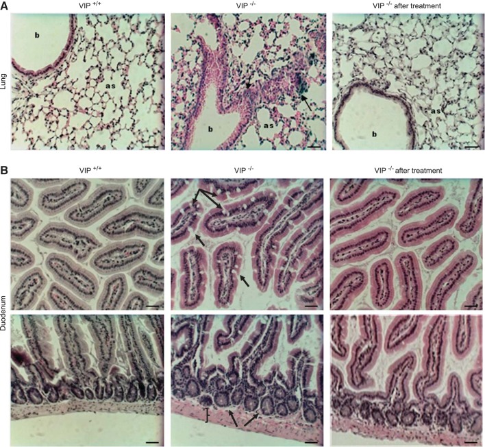
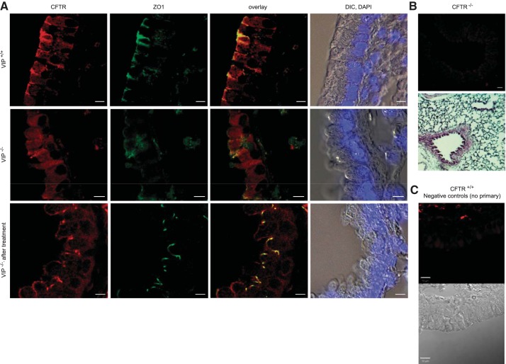
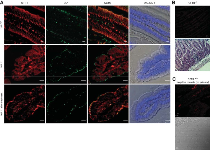
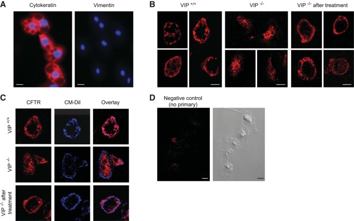
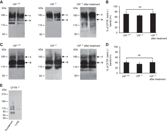
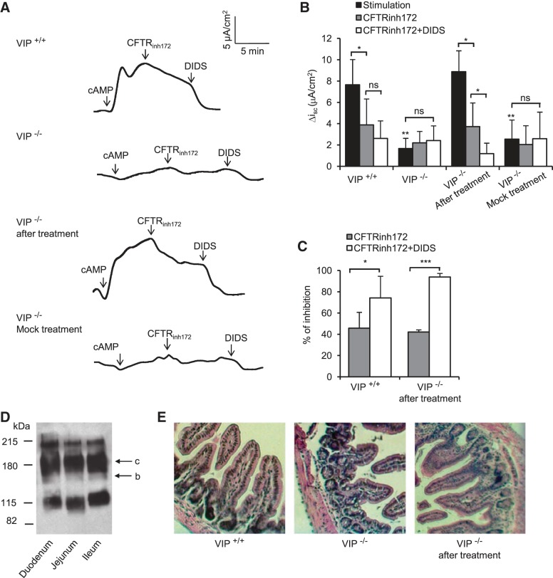
Vasoactive intestinal peptide (VIP), a neuropeptide, controls multiple functions in exocrine tissues, including inflammation, and relaxation of airway and vascular smooth muscles, and regulates CFTR-dependent secretion, which contributes to mucus hydration and local innate defense of the lung. We had previously reported that VIP stimulates the VPAC1 receptor, PKCϵ signaling cascade, and increases CFTR stability and function at the apical membrane of airway epithelial cells by reducing its internalization rate. Moreover, prolonged VIP stimulation corrects the molecular defects associated with F508del, the most common CFTR mutation responsible for the genetic disease cystic fibrosis. In the present study, we have examined the impact of the absence of VIP on CFTR maturation, cellular localization, and function in vivo using VIP knockout mice. We have conducted pathological assessments and detected signs of lung and intestinal disease. Immunodetection methods have shown that the absence of VIP results in CFTR intracellular retention despite normal expression and maturation levels. A subsequent loss of CFTR-dependent chloride current was measured in functional assays with Ussing chamber analysis of the small intestine ex vivo, creating a cystic fibrosis-like condition. Interestingly, intraperitoneal administration of VIP corrected tissue abnormalities, close to the wild-type phenotype, as well as associated defects in the vital CFTR protein. The results show in vivo a primary role for VIP chronic exposure in CFTR membrane stability and function and confirm in vitro data.

Keywords: CFTR; VIP; VIP-knockout mice; cystic fibrosis; epithelium.


Figures

Fig. 1.
Fig. 1.
Pathology of the lung and small intestine. Light microscopy pictures (20× objective) of hematoxylin and eosin (H and E)- stained tissue sections of the lung (A) and duodenum (B) of wild-type (VIP+/+), VIP-knockout (KO) (VIP−/−), and VIP-KO mice that received intraperitoneal injections of vasoactive intestinal peptide (VIP) for 3 wk (VIP−/− after treatment). A: compared with wild-type tissues, VIP-KO lungs show signs of inflammation (black arrows), inflammatory cells aggregation (dark blue staining), and thickening of the alveolar walls (as: alveolar sacs). b, Bronchioles. B: top panels show increased amount of goblet cells (black arrows) in the upper villi of VIP-KO duodenum. Crypts and muscle layer (bottom panels) are enlarged. Pathological signs observed in VIP-KO tissues were reversed by VIP administration. Scale bars = 50 μm. N = 5–7 mice in each group.
Fig. 2.
Fig. 2.
Apical localization of CFTR is compromised in VIP-KO lung and restored after VIP treatment. A: confocal microscopy images of CFTR immunofluorescent signals obtained from tissue sections of paraffin-embedded lungs from wild-type (VIP+/+), VIP-KO (VIP−/−), and VIP-KO mice that received intraperitoneal injections of VIP for 3 wk (VIP−/− after treatment). CFTR signal was mainly observed in the apical portion of the epithelium of wild-type tissues where it colocalizes with ZO1, but globally distributed in epithelial cells from VIP-KO tissues. CFTR surface expression was restored in tissues from VIP-KO mice that received VIP injections. Overlay signal from CFTR and ZO1 and differential interference contrast (DIC) pictures with cell nuclei stained with DAPI are also presented. B and C: negative controls for CFTR immunostaining. B, top panels: representative image of CFTR-KO (C57Bl/6 cftr−/−) lung tissue immunostained with the CFTR monoclonal antibody (MAB1660), revealed with a goat anti-mouse secondary antibody coupled to Cy3 as in A. H and E-stained section is also shown. C: negative control for wild-type CFTR (CFTR+/+) immunostaining of the lung in which the primary antibody was omitted. Corresponding DIC picture is shown. Scale bars: 5 μm. N = 5–7 mice in each group.
Fig. 3.
Fig. 3.
Apical localization of CFTR is compromised in VIP-KO duodenum and restored after VIP treatment. A: confocal microscopy images of CFTR immunofluorescent signals obtained from tissue sections of paraffin-embedded duodenum from wild-type (VIP+/+), VIP-KO (VIP−/−), and VIP-KO mice that received intraperitoneal injections of VIP for 3 wk (VIP−/− after treatment). CFTR signal was mainly observed in the apical portion of the epithelium of wild-type tissues where it colocalizes with ZO1, but globally distributed in epithelial cells from VIP-KO tissues. CFTR surface expression was restored in tissues from VIP-KO mice that received VIP injections. Overlay signal from CFTR and ZO1 and DIC pictures with cell nuclei stained with DAPI are also presented. B and C: negative controls for CFTR immunostaining. B, top panels: representative image of CFTR-KO (C57Bl/6 cftr−/−) duodenum tissue immunostained with the CFTR monoclonal antibody (MAB1660), revealed with a goat anti-mouse secondary antibody coupled to Cy3 as in A. H and E-stained section is also shown. C: negative control for wild-type CFTR (CFTR+/+) immunostaining of the duodenum in which the primary antibody was omitted. Corresponding DIC picture is shown. Scale bars: 5 μm. N = 5–7 mice in each group.
Fig. 4.
Fig. 4.
CFTR localization in primary tracheal epithelial cells. A: cytokeratin and vimentin immunostainings (red) were performed on epithelial cells extracted by enzymatic digestion from mice trachea to confirm the epithelial phenotype of extracted cells. DAPI was used to stain nuclei (blue). Scale bar = 20 μm. B: tracheal epithelial cells were fixed and permeabilized before immunostaining with the MAB1660 anti-CFTR antibody, followed by a goat anti-mouse secondary antibody coupled to Cy3. CFTR immunofluorescent signal was mainly found at the membrane of epithelial cells extracted from wild-type mice tracheas (VIP+/+), intracellularly in VIP-KO cells (VIP−/−), and at the surface of epithelial cells from VIP-KO mice that received VIP injections (VIP−/− after treatment). Scale bars = 10 μm. C: tracheal epithelial cells were labeled with the membrane dye CM-Dil (blue signal) followed by immunostaining for CFTR (red signal) as in B. D: negative control in which the primary antibody was omitted. Corresponding DIC picture is shown. Epithelial cells were extracted from 10 mice in each group and immunostaining repeated 3 times.
Fig. 5.
Fig. 5.
CFTR expression and maturation. A and C: representative immunoblots showing similar CFTR expression and maturation level in tissue lysates from duodenum (A) and lung (C), from two different mice in each group: wild-type (VIP+/+), VIP-KO (VIP−/−), and VIP-KO mice that received intraperitoneal injections of VIP for 3 wk (VIP−/− after treatment). Mature fully glycosylated band c and immature core-glycosylated band b are indicated by arrows. B and D: histograms showing average values from densitometry measurements of CFTR mature band c (∼180 kDa) expressed as percentage (%) of total CFTR (bands b + c) in duodenum (B) and lung (D) of wild-type, VIP-KO, and VIP-KO mice treated with VIP. ns, not statistically significant. E: negative control immunoblot showing the absence of CFTR signal in tissue lysates from CFTR-KO duodenum and CFTR-KO lung as indicated. Tissue lysates from 5 mice in each group were analyzed and used to calculate average densitometry ratios.
Fig. 6.
Fig. 6.
CFTR-dependent chloride secretion. A: short-circuit current, measured in voltage-clamp mode, generated by ileums from wild-type (VIP+/+), VIP-KO (VIP−/−), and VIP-KO mice that received intraperitoneal injections of VIP for 3 wk (VIP−/− after VIP treatment) or PBS (VIP−/− mock treatment). Ileums were mounted in Ussing chambers with an aperture size of 0.125 cm2. Krebs buffer was placed on the blood side and a chloride-free buffer on the luminal side. After allowing the tissue to stabilize for ∼10 min, CFTR was stimulated by a cAMP cocktail [10 μM forskolin + 1 mM 3-isobutyl-1-methylxanthine (IBMX) + 200 μM 8-(4-chlorophenylthio)-2′-O-methyladenosine 3′,5′-cyclic monophosphate monosodium hydrate (cpt-cAMP)] added to the lumen side. The selective CFTR inhibitor CFTRinh172 (20 μM) was added ∼5–10 min after stimulation after reaching a plateau of activation to unmask CFTR-dependent chloride current, and the nonspecific chloride channel blocker 4,4′-diisothiocyanatostilbene-2,2′-disulfonic acid disodium salt hydrate (DIDS, 200 μM) was subsequently added to inhibit all other chloride channel activity. CFTR-dependent chloride current was observed in wild-type ileum (VIP+/+) but absent from VIP-KO tissues (VIP−/−). The bottom traces show functional recovery after VIP treatment, but not with negative control injections of PBS only. B: histogram showing average values of short-circuit current differences between baseline and plateau of stimulation or inhibition (ΔIsc). C: histogram showing average values of the percentage of inhibition of the cAMP-stimulated Disc current by the CFTRinh172 and after the additive action of CFTRinh172 and DIDS. Asterisks above black bars indicate a significant difference with stimulated ΔIsc measured in wild-type ileum. D: immunoblot showing similar expression level of CFTR in duodenum, jejunum, and ileum. Bands c (mature fully glycosylated) and b (immature core-glycosylated) are indicated by arrow. E: histology sections of corresponding ileums as indicated, showing same pathology as duodenum from VIP-KO mice, and recovery after VIP injections. Ileums from 5 different mice in each group were used. *P < 0.05, **P < 0.01, ***P < 0.001.
Fig. 7.
Fig. 7.
Detection of VIP receptors expression by RT-PCR. VPAC1, VPAC2, and PAC1 receptor expression was detected by RT-PCR after RNA extraction from whole tissue lysates. PCR products were subjected to 0.8% agarose gel electrophoresis. Left lane: φx DNA ladder. Expected sizes (bp) of amplified products are indicated. A and B: samples from lungs (A) and duodenum (B) of wild-type (VIP+/+), VIP-KO (VIP−/−), and VIP-KO mice that received intraperitoneal injections of VIP for 3 wk (VIP−/− after treatment) were amplified with specific primers for each receptor (see materials and methods). C: positive control using PKCα. D: negative control which had cDNA excluded from the reaction. E: CFTR, VPAC1, VPAC2, and PAC1 receptor expression in epithelial cells extracted from wild-type mice tracheas. PCR products were subjected to 1.5% agarose gel electrophoresis. mRNA extracted from primary cultures of epithelial cells were amplified with specific primers (see materials and methods). PKCα was used as positive control. Reaction in which cDNA was excluded was used a negative control.
Fig. 8.
Fig. 8.
VIP receptor overexpression in VIP-KO mice. A and B: representative immunoblots showing expression of VPAC1, VPAC2, and PAC1 receptors in tissue lysates from lung (A) and duodenum (B) of wild-type (VIP+/+), VIP-KO (VIP−/−), and VIP-KO mice that received intraperitoneal injections of VIP for 3 wk (VIP−/− after treatment). Expression levels in lysates from two different mice in each group are presented. C–E: changes in each receptor's expression were calculated from densitometry measurement of scanned immunoblots and reported as percentage of corresponding actin signal. Values are means ± SE for N = 5. **P < 0.01, ***P < 0.001. ns, not statically significant.

References

    1. Alcolado N, Conrad DJ, Rafferty S, Chappe FG, Chappe VM. VIP-dependent increase in F508del-CFTR membrane localization is mediated by PKCε. Am J Physiol Cell Physiol 301: C53–C65, 2011 - PubMed
    1. Alessandrini F, Thakkar M, Foda HD, Said SI, Lodi R, Pakbaz H, Schraufnagel DE. Vasoactive intestinal peptide enhances lung preservation. Transplantation 56: 964–973, 1993 - PubMed
    1. Ameen NA, Ardito T, Kashgarian M, Marino CR. A unique subset of rat and human intestinal villus cells express the cystic fibrosis transmembrane conductance regulator. Gastroenterology 108: 1016–1023, 1995 - PubMed
    1. Ameen NA, Martensson B, Bourguinon L, Marino C, Isenberg J, McLaughlin GE. CFTR channel insertion to the apical surface in rat duodenal villus epithelial cells is upregulated by VIP in vivo. J Cell Sci 112: 887–894, 1999 - PubMed
    1. Ameen NA, van Donselaar E, Posthuma G, de Jonge H, McLaughlin G, Geuze HJ, Marino C, Peters PJ. Subcellular distribution of CFTR in rat intestine supports a physiologic role for CFTR regulation by vesicle traffic. Histochem Cell Biol 114: 219–228, 2000 - PubMed

Publication types

MeSH terms

Substances

LinkOut - more resources