. 2014 Aug 1;307(3):C266-77.
doi: 10.1152/ajpcell.00135.2014. Epub 2014 Jun 4.

Chronic opioids regulate KATP channel subunit Kir6.2 and carbonic anhydrase I and II expression in rat adrenal chromaffin cells via HIF-2α and protein kinase A

Affiliations

Chronic opioids regulate KATP channel subunit Kir6.2 and carbonic anhydrase I and II expression in rat adrenal chromaffin cells via HIF-2α and protein kinase A

Shaima Salman et al. Am J Physiol Cell Physiol. .

Abstract

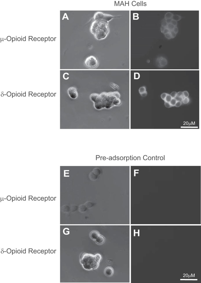
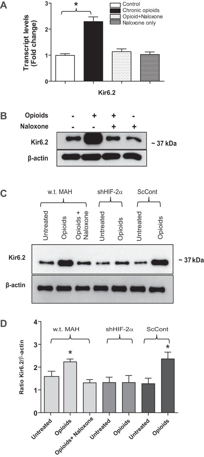
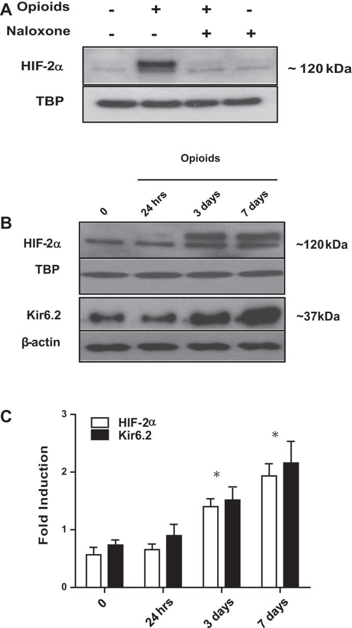
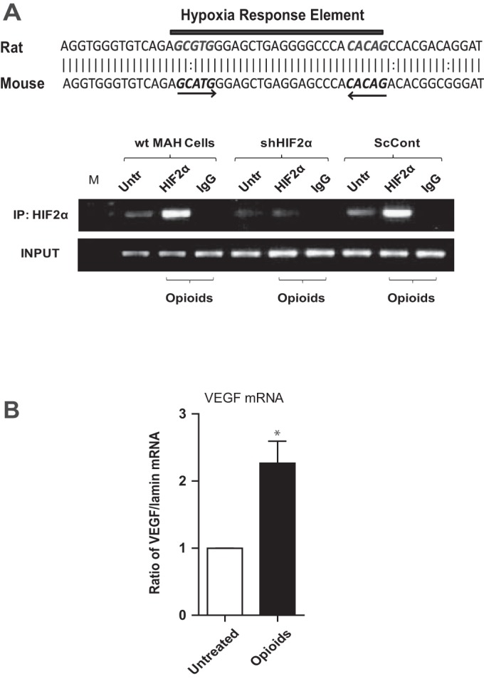
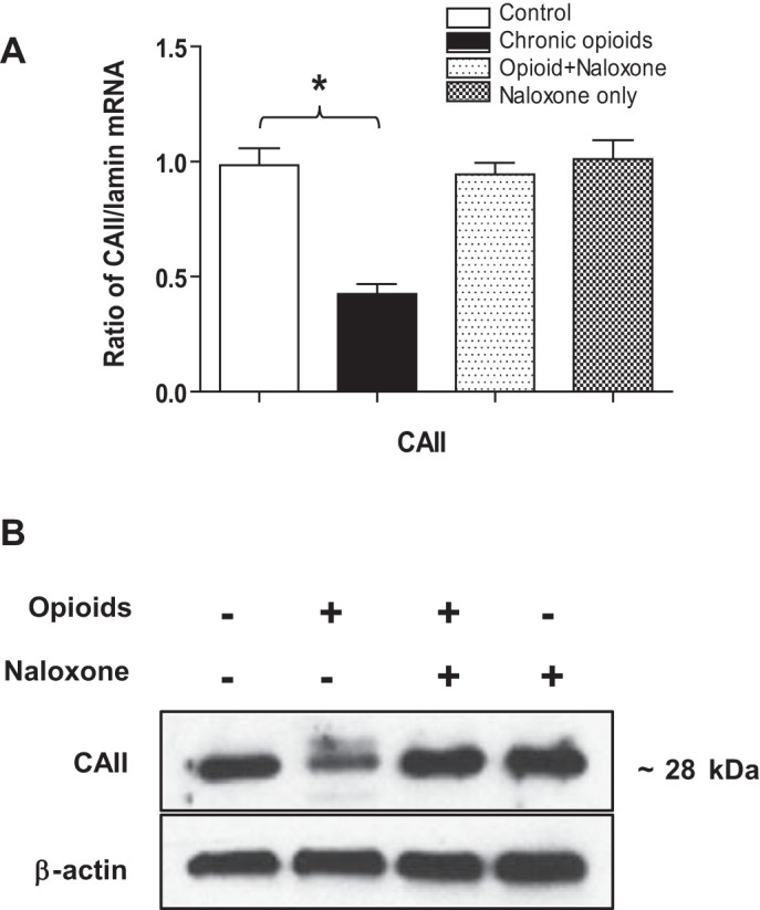
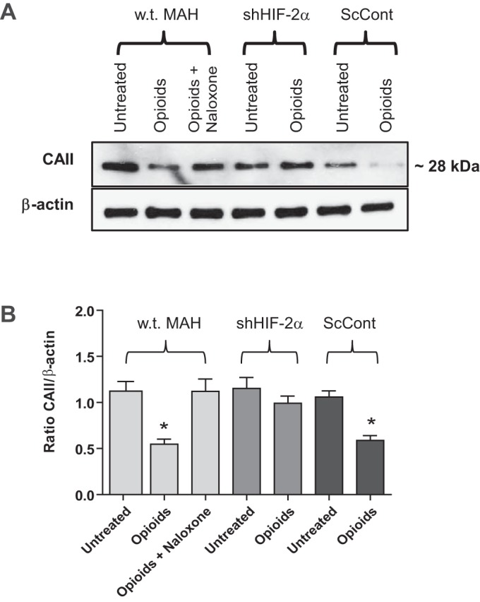
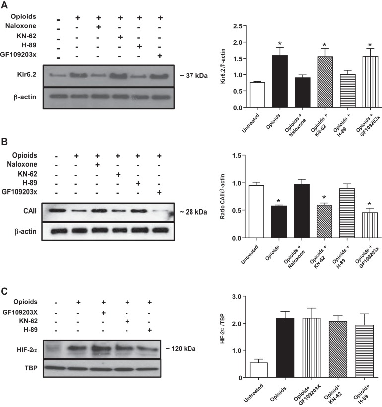
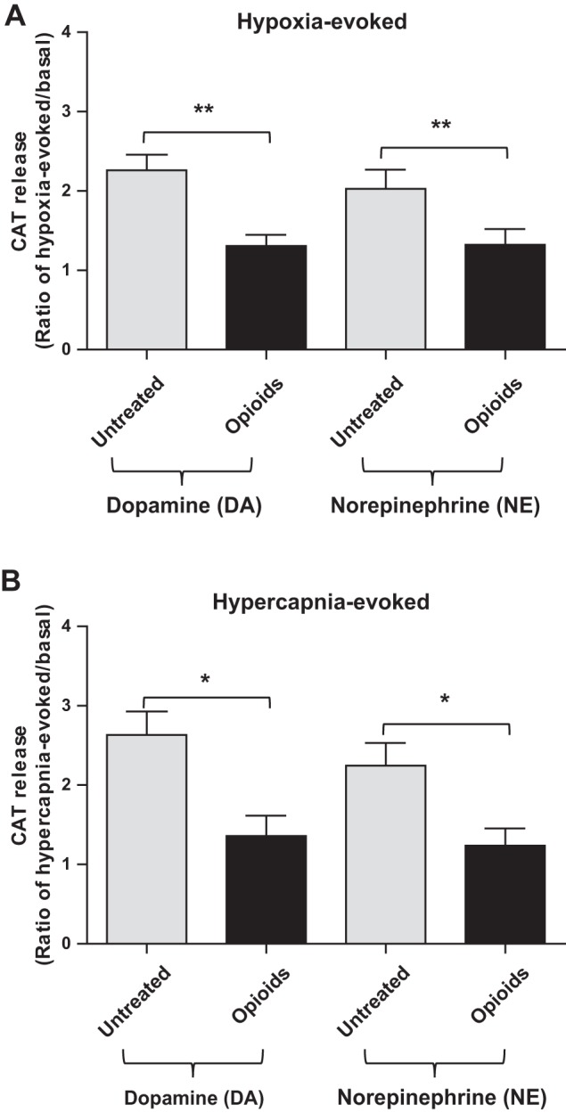
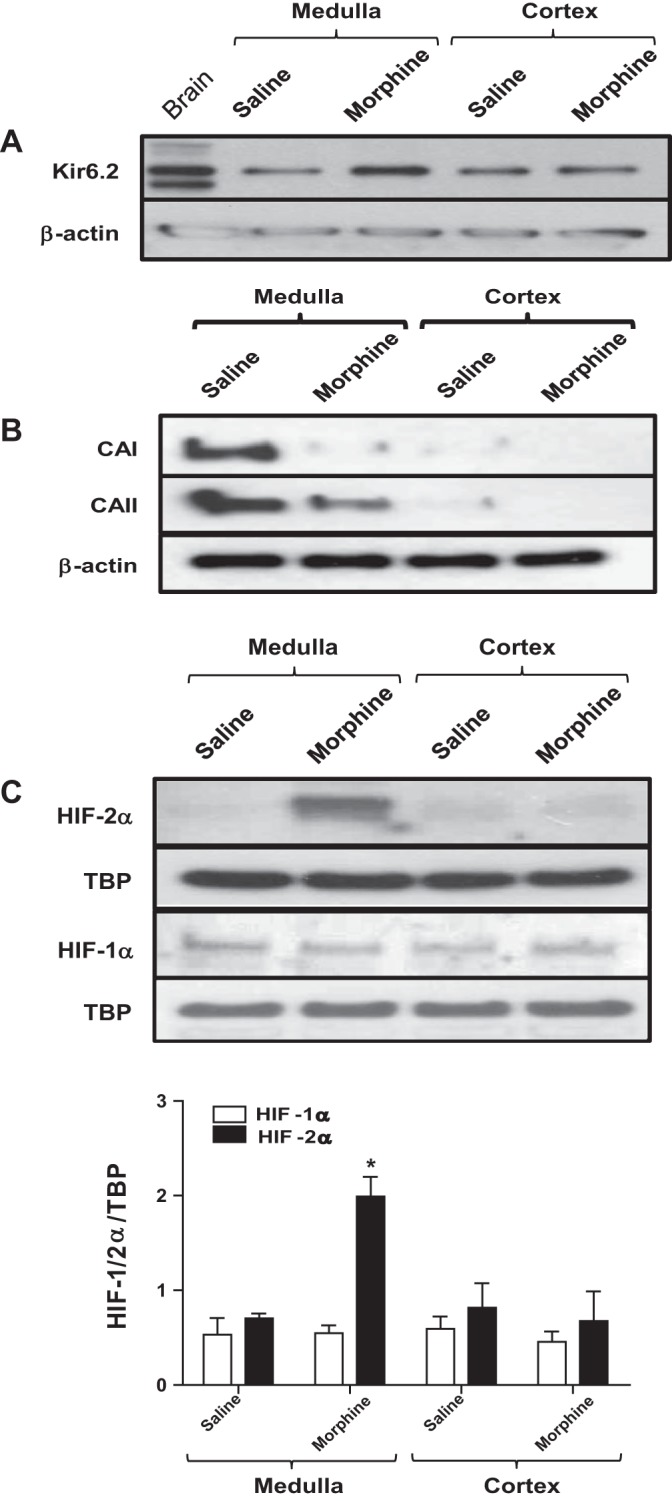
At birth, asphyxial stressors such as hypoxia and hypercapnia are important physiological stimuli for adrenal catecholamine release that is critical for the proper transition to extrauterine life. We recently showed that chronic opioids blunt chemosensitivity of neonatal rat adrenomedullary chromaffin cells (AMCs) to hypoxia and hypercapnia. This blunting was attributable to increased ATP-sensitive K(+) (KATP) channel and decreased carbonic anhydrase (CA) I and II expression, respectively, and involved μ- and δ-opioid receptor signaling pathways. To address underlying molecular mechanisms, we first exposed an O2- and CO2-sensitive, immortalized rat chromaffin cell line (MAH cells) to combined μ {[d-Arg(2),Ly(4)]dermorphin-(1-4)-amide}- and δ ([d-Pen(2),5,P-Cl-Phe(4)]enkephalin)-opioid agonists (2 μM) for ∼7 days. Western blot and quantitative real-time PCR analysis revealed that chronic opioids increased KATP channel subunit Kir6.2 and decreased CAII expression; both effects were blocked by naloxone and were absent in hypoxia-inducible factor (HIF)-2α-deficient MAH cells. Chronic opioids also stimulated HIF-2α accumulation along a time course similar to Kir6.2. Chromatin immunoprecipitation assays on opioid-treated cells revealed the binding of HIF-2α to a hypoxia response element in the promoter region of the Kir6.2 gene. The opioid-induced regulation of Kir6.2 and CAII was dependent on protein kinase A, but not protein kinase C or calmodulin kinase, activity. Interestingly, a similar pattern of HIF-2α, Kir6.2, and CAII regulation (including downregulation of CAI) was replicated in chromaffin tissue obtained from rat pups born to dams exposed to morphine throughout gestation. Collectively, these data reveal novel mechanisms by which chronic opioids blunt asphyxial chemosensitivity in AMCs, thereby contributing to abnormal arousal responses in the offspring of opiate-addicted mothers.

Keywords: adenosine 5′-triphosphate-sensitive potassium channels; carbonic anhydrase; hypoxia-inducible factor-2α; opioid.


Figures

Fig. 1.
Fig. 1.
Immunofluorescence staining of μ- and δ-opioid receptors in MAH cell cultures. Corresponding phase-contrast (top left) and fluorescence (top right) images of MAH cells showing positive immunostaining for μ-opioid (A and B) and δ-opioid (C and D) receptors. In control experiments for μ-opioid (E and F) and δ-opioid (G and H) receptor, the primary antibody was preincubated with excess antigen before application to the cells, followed by FITC-conjugated secondary antibody.
Fig. 2.
Fig. 2.
Quantitative real-time PCR (QPCR) and Western blot analyses of ATP-sensitive K+ (KATP) channel subunit Kir6.2 expression in MAH cells. A: QPCR analysis of Kir6.2 mRNA in control MAH cells, MAH cells cultured with combined μ- and δ- opioid agonists (2 μM) ± naloxone (2 μM), and MAH cells cultured with naloxone for 7 days. Note significant upregulation of Kir6.2 transcript in opioid-treated cells (*P < 0.05; n = 4 for each group); one-way ANOVA was used for multiple comparisons within groups. B: Western blot analysis showing increased expression of Kir6.2 protein in opioid-treated MAH cells and its prevention in the presence of naloxone; β-actin was used as an internal control (n = 3). Western blot analyses of Kir6.2 subunit expression in wild-type (wt), hypoxia inducible factor-2α (HIF-2α)-deficient (shHIF-2α), and scrambled control (ScCont) MAH cells cultured with combined opioid agonists (2 μM) or in the presence of naloxone (2 μM) for 7 days (C). Note absence of Kir6.2 subunit upregulation in shHIF-2α MAH cells. Densitometric analysis of changes in Kir6.2 subunit expression normalized β-actin (D); n = 3 for each group, *P < 0.05.
Fig. 3.
Fig. 3.
Effects of chronic opioid exposure on Kir6.2 and HIF-2α expression in MAH cells. A: Western blot analysis of HIF-2α accumulation in MAH cells cultured with combined μ- and δ- opioid agonists (2 μM) ± naloxone (2 μM), or with naloxone only (2 μM), for 7 days. B: time-dependent HIF-2α and Kir6.2 protein expression in MAH cells exposed to combined opioids (2 μM) for 24 h, 3 days, and 7 days in culture. β-Actin was used as loading control for cytoplasmic extracts in the case of Kir6.2 and TATA-binding protein (TBP) for nuclear extracts in the case of HIF-2α. Results show progressive increase in Kir6.2 expression that parallels HIF-2α accumulation following chronic opioids; for both Kir6.2 and HIF-2α, the increase is significant at 3 and 7 days (B and C). C: densitometric analysis demonstrating the fold induction in Kir6.2 subunit expression and HIF-2α accumulation normalized to loading control. Data are expressed as means ± SE for three independent experiments for each group, *P < 0.05.
Fig. 4.
Fig. 4.
Chromatin immunoprecipitation (ChIP) assay demonstrating binding of HIF-2α to the promoter region of Kir6.2 gene in opioid-treated MAH cells. Note hypoxia response element (HRE) within the promoter region of rat and mouse Kir6.2 gene (A, top); the HIF core site (GCGTG) spans nucleotides −1087 to −1083 and HIF ancillary site (CACAG) spans nucleotides −1065 to −1061. Lysates from untreated control (Untr) and opioid-treated wt, shHIF2α, and ScCont MAH cells were subjected to ChIP assay using a HIF-2α polyclonal antibody (A, bottom). PCR analysis was performed using a primer pair designed to span the putative HRE upstream of the promoter region. Technical controls include a ChIP performed using nonspecific IgG monoclonal antibody (IgG) and a starting material control (Input). B: vascular endothelial growth factor (VEGF) mRNA expression as determined by QPCR analysis in control (untreated) and opioid-treated MAH cells (n = 3). Data are expressed as means ± SE for three independent experiments for each group, *P < 0.05.
Fig. 5.
Fig. 5.
Effects of chronic opioid exposure on carbonic anhydrase II (CAII) expression in MAH cells. A: mRNA expression analysis of CAII in control MAH cells, in MAH cells cultured with combined μ- and δ-opioid agonists (2 μM) ± naloxone (2 μM), and in MAH cells cultured with naloxone (2 μM) only for 7 days. *P < 0.05, n = 3 for each group. One-way ANOVA was used for multiple comparisons within groups. B: Western blot analysis showing expression of CAII protein in MAH cells grown under similar conditions. β-Actin was used as an internal control. Note coincubation with naloxone prevented the chronic opioid-induced decrease in CAII expression in A and B.
Fig. 6.
Fig. 6.
Effects of HIF-2α-deficiency on CAII expression in opioid-treated MAH cells. A, top: Western blots of CAII protein expression in control MAH cells, shHIF2α MAH cells, and ScCont MAH cells, cultured with or without combined opioid agonists (2 μM) or in the presence of naloxone (2 μM) for 7 days. B: densitometric analysis of CAII protein expression normalized to β-actin under the various conditions indicated. Note the opioid-mediated downregulation of CAII is absent in HIF-2α-deficient MAH cells. Data are expressed as means ± SE for three independent experiments for each group, *P < 0.05.
Fig. 7.
Fig. 7.
Effects of protein kinase inhibitors on expression of Kir6.2, CAII, and HIF-2α in MAH cells following chronic opioid exposure. A: Western blot analysis showing the effects of protein kinase inhibition on Kir6.2 expression in opioid-treated MAH cells. Inhibitors of protein kinase A (PKA) (H-89) (2 μM), but not inhibitors of protein kinase C (GF-109203X) (2 μM) or calmodulin (CaM) kinase (KN-62) (3 μM), prevented the upregulation of Kir6.2 in opioid-treated cells. Densitometric analysis of Kir6.2 subunit expression normalized to β-actin is shown in histogram (right). A similar protocol was used to study the effects of protein kinase inhibitors on CAII expression relative to β-actin (B), and HIF-2α accumulation relative to TBP (C), in opioid-treated MAH cells. Note the PKA inhibitor (H-89) prevented the opioid-induced downregulation of CAII expression, but not HIF-2α accumulation, in MAH cells. Data are expressed as means ± SE for three independent experiments for each group, *P < 0.05.
Fig. 8.
Fig. 8.
Effects of chronic opioids on hypoxia- and hypercapnia-evoked dopamine (DA) and norepinephrine (NE) release from MAH cells. ELISA was used to measure DA and NE release, and data were expressed as a ratio of evoked relative to basal release. Chemostimulation with hypoxia (2% O2; 15 min) (A) and isohydric hypercapnia (10% CO2; pH 7.4) (B) caused a significant increase in DA and NE release in control (untreated) MAH cells. Following exposure of MAH cells to chronic opioids for ∼1 wk in vitro, the effect of hypoxia and hypercapnia on DA and NE release was significantly reduced. Data are represented as means ± SE, where n = 5 for A and 4 for B (*P < 0.05 and **P < 0.01).
Fig. 9.
Fig. 9.
Effects of maternal morphine injections on the expression of KATP channel subunit (Kir6.2), carbonic anhydrase I (CAI) and CAII enzymes, and HIFs in adrenal gland tissues of affected neonates. Western blot analyses of KATP channel subunit Kir6.2 (A), CAI and -II (B), and HIF-1α and -2α (C) expression in adrenal medulla (AM) and adrenal cortex (AC) of saline- and morphine-exposed rat pups. Note the increased Kir6.2 subunit and reduced CAI and CAII expression (relative to β-actin) in AM, but not AC, of morphine-exposed pups. Also, note the selective increase in HIF-2α, but not HIF-1α, accumulation (relative to TBP) in AM of morphine-exposed pups as shown in C. Data are expressed as means ± SE for three independent experiments for each group, *P < 0.05.

Similar articles

Cited by

References

    1. Bishop T, Gallagher D, Pascual A, Lygate CA, de Bono JP, Nicholls LG, Ortega-Saenz P, Oster H, Wijeyekoon B, Sutherland AI, Grosfeld A, Aragones J, Schneider M, van Geyte K, Teixeira D, Diez-Juan A, Lopez-Barneo J, Channon KM, Maxwell PH, Pugh CW, Davies AM, Carmeliet P, Ratcliffe PJ. Abnormal sympathoadrenal development and systemic hypotension in PHD3−/− mice. Mol Cell Biol 28: 3386–3400, 2008 - PMC - PubMed
    1. Brown ST, Kelly KF, Daniel JM, Nurse CA. Hypoxia inducible factor (HIF)-2 alpha is required for the development of the catecholaminergic phenotype of sympathoadrenal cells. J Neurochem 110: 622–630, 2009 - PubMed
    1. Brown ST, Nurse CA. Induction of HIF-2alpha is dependent on mitochondrial O2 consumption in an O2-sensitive adrenomedullary chromaffin cell line. Am J Physiol Cell Physiol 294: C1305–C1312, 2008 - PubMed
    1. Burns L, Conroy E, Mattick RP. Infant mortality among women on a methadone program during pregnancy. Drug Alcohol Rev 29: 551–556, 2010 - PubMed
    1. Buttigieg J, Brown S, Holloway AC, Nurse CA. Chronic nicotine blunts hypoxic sensitivity in perinatal rat adrenal chromaffin cells via upregulation of KATP channels: role of alpha7 nicotinic acetylcholine receptor and hypoxia-inducible factor-2alpha. J Neurosci 29: 7137–7147, 2009 - PMC - PubMed

Publication types

MeSH terms

Substances

LinkOut - more resources