Review
. 2014 Jun 4;6(239):239sr1.
doi: 10.1126/scitranslmed.3005101.

Imaging and nanomedicine in inflammatory atherosclerosis

Affiliations
Review

Imaging and nanomedicine in inflammatory atherosclerosis

Willem J M Mulder et al. Sci Transl Med. .

Abstract

Bioengineering provides unique opportunities to better understand and manage atherosclerotic disease. The field is entering a new era that merges the latest biological insights into inflammatory disease processes with targeted imaging and nanomedicine. Preclinical cardiovascular molecular imaging allows the in vivo study of targeted nanotherapeutics specifically directed toward immune system components that drive atherosclerotic plaque development and complication. The first multicenter trials highlight the potential contribution of multimodality imaging to more efficient drug development. This review describes how the integration of engineering, nanotechnology, and cardiovascular immunology may yield precision diagnostics and efficient therapeutics for atherosclerosis and its ischemic complications.


Figures

Fig. 1
Fig. 1. Imaging cellular processes and inflammation in atherosclerotic plaques
Inflammatory atherosclerosis can be diagnosed and monitored by clinical and preclinical imaging methods, some of which require the administration of targeted agents. Preclinically, key inflammation imaging techniques include macrophage imaging in the vessel wall with nanoparticles, adhesion molecule imaging, or protease-specific agents, whereas splenic monocytes that migrate to the plaque can be monitored by intravital microscopy. In a clinical setting, MRI can visualize macrophage burden in human plaques, whereas dynamic contrast-enhanced MRI (DCE-MRI) and FDG-PET/CT allow the quantification of plaque neovessel permeability and inflammation, respectively. Lowercase roman numerals match imaging approach to location in the atherosclerotic vessel. Image in (iii) obtained from (35) with permission; in (iv), from (90) with permission; in (v), from (53) with permission. Image in (vi) is an original image from Z.A.F.
Fig. 2
Fig. 2. Nanoparticle imaging and corresponding diagnostic modalities
Nanoparticles can be labeled with a variety of imaging agents to enable their detection in plaque cells with CT, MRI, optical methods, or nuclear imaging, such as PET and SPECT. The relative costs, sensitivity, scan time range, and resolution range are indicated. CT image obtained from (47) with permission; MRI image from (52) with permission; optical image from (35) with permission; PET image from (55) with permission. DTPA, diethylenetriamine pentaacetic acid; DOTA, 1,4,7,10-tetraazacyclododecane-1,4,7,10-tetraacetic acid.
Fig. 3
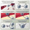
Fig. 3. Therapeutic targets in inflammatory atherosclerosis
A cascade of processes that contribute to macrophage activity and inflammation in atherosclerotic plaques can be specifically targeted to halt the progression of the disease. The migration of monocytes from the bone marrow or spleen to atherosclerotic plaque can be inhibited; the phenotype of monocytes can be modulated; monocyte migration into the plaque can be halted, or monocytes/macrophages departing the plaque can be stimulated. Moreover, macrophage proliferation and the excretion of harmful proteases or cytokines are compelling therapeutic targets.
Fig. 4
Fig. 4. Considerations for an imaging-, nanotherapy-, and immunology-facilitated atherosclerosis preclinical study
The early-stage development of an anti-inflammatory nanotherapy for atherosclerosis requires comprehensive evaluation of various pharmacological aspects and effects. These include the determination of the biodistribution (by imaging), toxicology, and therapeutic mechanism. Nanotherapy formulations should consider the target, delivery mechanism, and what type of drug will be included. Preclinically, efficacy of the nanotherapy and the immunological effects can be determined commonly in a mouse or rabbit model of atherosclerosis. Immunohistochemistry image obtained from (35) with permission. Plaque profiling images obtained from (52) with permission. Molecular imaging panel obtained from (96) with permission. Vessel wall images obtained from (52) with permission.

References

    1. Moore KJ, Tabas I. Macrophages in the pathogenesis of atherosclerosis. Cell. 2011;145:341–355. - PMC - PubMed
    1. Libby P, Ridker PM, Hansson GK. Progress and challenges in translating the biology of atherosclerosis. Nature. 2011;473:317–325. - PubMed
    1. Swirski FK, Nahrendorf M. Leukocyte behavior in atherosclerosis, myocardial infarction, and heart failure. Science. 2013;339:161–166. - PMC - PubMed
    1. Libby P. Inflammation in atherosclerosis. Nature. 2002;420:868–874. - PubMed
    1. Mitka M. New basic care goals seek to rein in global rise in cardiovascular disease. JAMA. 2012;308:1725–1726. - PubMed

Publication types