Automated detection of cortical dysplasia type II in MRI-negative epilepsy
- PMID: 24898923
- PMCID: PMC4114179
- DOI: 10.1212/WNL.0000000000000543
Automated detection of cortical dysplasia type II in MRI-negative epilepsy
Abstract
Objective: To detect automatically focal cortical dysplasia (FCD) type II in patients with extratemporal epilepsy initially diagnosed as MRI-negative on routine inspection of 1.5 and 3.0T scans.
Methods: We implemented an automated classifier relying on surface-based features of FCD morphology and intensity, taking advantage of their covariance. The method was tested on 19 patients (15 with histologically confirmed FCD) scanned at 3.0T, and cross-validated using a leave-one-out strategy. We assessed specificity in 24 healthy controls and 11 disease controls with temporal lobe epilepsy. Cross-dataset classification performance was evaluated in 20 healthy controls and 14 patients with histologically verified FCD examined at 1.5T.
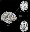
Results: Sensitivity was 74%, with 100% specificity (i.e., no lesions detected in healthy or disease controls). In 50% of cases, a single cluster colocalized with the FCD lesion, while in the remaining cases a median of 1 extralesional cluster was found. Applying the classifier (trained on 3.0T data) to the 1.5T dataset yielded comparable performance (sensitivity 71%, specificity 95%).
Conclusion: In patients initially diagnosed as MRI-negative, our fully automated multivariate approach offered a substantial gain in sensitivity over standard radiologic assessment. The proposed method showed generalizability across cohorts, scanners, and field strengths. Machine learning may assist presurgical decision-making by facilitating hypothesis formulation about the epileptogenic zone.
Classification of evidence: This study provides Class II evidence that automated machine learning of MRI patterns accurately identifies FCD among patients with extratemporal epilepsy initially diagnosed as MRI-negative.
© 2014 American Academy of Neurology.
Figures


References
-
- Sisodiya SM, Fauser S, Cross JH, Thom M. Focal cortical dysplasia type II: biological features and clinical perspectives. Lancet Neurol 2009;8:830–843 - PubMed
-
- Lerner JT, Salamon N, Hauptman JS, et al. Assessment and surgical outcomes for mild type I and severe type II cortical dysplasia: a critical review and the UCLA experience. Epilepsia 2009;50:1310–1335 - PubMed
-
- Muhlebner A, Coras R, Kobow K, et al. Neuropathologic measurements in focal cortical dysplasias: validation of the ILAE 2011 classification system and diagnostic implications for MRI. Acta Neuropathol 2012;123:259–272 - PubMed
Publication types
MeSH terms
Grants and funding
LinkOut - more resources
Full Text Sources
Other Literature Sources
Medical
Research Materials
