. 2014 Aug;88(16):9049-59.
doi: 10.1128/JVI.01161-14. Epub 2014 Jun 4.

Modification of the hepatitis B virus envelope protein glycosylation pattern interferes with secretion of viral particles, infectivity, and susceptibility to neutralizing antibodies

Affiliations

Modification of the hepatitis B virus envelope protein glycosylation pattern interferes with secretion of viral particles, infectivity, and susceptibility to neutralizing antibodies

Romain Julithe et al. J Virol. 2014 Aug.

Abstract

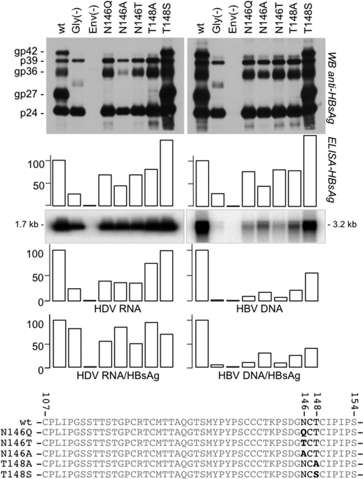
The envelope proteins of hepatitis B virus (HBV) bear an N-linked glycosylation site at N146 within the immunodominant a-determinant in the antigenic loop (AGL) region. This glycosylation site is never fully functional, leading to a nearly 1/1 ratio of glycosylated/nonglycosylated isoforms in the viral envelope. Here we investigated the requirement for a precise positioning of N-linked glycan at amino acid 146 and the functions associated with the glycosylated and nonglycosylated isoforms. We observed that the removal of the N146 glycosylation site by mutagenesis was permissive to envelope protein synthesis and stability and to secretion of subviral particles (SVPs) and hepatitis delta virus (HDV) virions, but it was detrimental to HBV virion production. Several positions in the AGL could substitute for position 146 as the glycosylation acceptor site. At position 146, neither a glycan chain nor asparagine was absolutely required for infectivity, but there was a preference for a polar residue. Envelope proteins bearing 5 AGL glycosylation sites became hyperglycosylated, leading to an increased capacity for SVP secretion at the expense of HBV and HDV virion secretion. Infectivity-compatible N-glycosylation sites could be inserted at 3 positions (positions 115, 129, and 136), but when all three positions were glycosylated, the hyperglycosylated mutant was substantially attenuated at viral entry, while it acquired resistance to neutralizing antibodies. Taken together, these findings suggest that the nonglycosylated N146 is essential for infectivity, while the glycosylated form, in addition to its importance for HBV virion secretion, is instrumental in shielding the a-determinant from neutralizing antibodies.

Importance: At the surface of HBV particles, the immunodominant a-determinant is the main target of neutralizing antibodies and an essential determinant of infectivity. It contains an N-glycosylation site at position 146, which is functional on only half of the envelope proteins. Our data suggest that the coexistence of nonglycosylated and glycosylated N146 at the surface of HBV reflects the dual function of this determinant in infectivity and immune escape. Hence, a modification of the HBV glycosylation pattern affects not only virion assembly and infectivity but also immune escape.


Figures

FIG 1
FIG 1
HBV envelope proteins and their glycosylation patterns. (A) The secondary structure model for the HBV envelope proteins at the viral membrane (mbne), including N-linked glycans (closed circles) in the antigenic loop (AGL) and pre-S2 domain and an O-linked glycans (open circle) in the pre-S2 domain, is presented. (B) HBV envelope proteins derived from SVPs produced in Huh-7 cells were subjected to immunoblot analysis using a rabbit R247 anti-S antibody. Note that HBV envelope proteins of genotype A (adw2 subtype) and genotype D (ayw3 subtype) bear one and two glycosylation sites in the pre-S2 domain, respectively. The glycosylated (gp) and nonglycosylated (p) forms of S-HBsAg (S), M-HBsAg (M), and L-HBsAg (L) proteins are indicated. Note that the gggpM and ggpM isoforms of M-HBsAg genotype D ayw3 comigrate as gp36 in SDS-polyacrylamide gels. (C) Schematic representation of the different isoforms of the HBV genotype D envelope proteins (ayw3 subtype). (D) Schematic representation of the genotype A M-HBsAg protein (adw2 subtype).
FIG 2
FIG 2
Synthesis and secretion of wt and mutant (N146T) HBV envelope proteins. Two days after transfection of Huh-7 cells with plasmid DNA carrying the sequences for wt or N146T mutant HBV envelope proteins, cells were pulse-labeled with [35S]Cys for 1 h, and labeling was chased for the indicated time period before harvesting (22). (Top and middle) Labeled proteins from cell lysates (lanes C) and culture medium (lanes M) were immunoprecipitated with rabbit R247 anti-S antibodies and analyzed by SDS-PAGE. After electrophoresis, the gel was dried and subjected to fluorography at −70°C for 48 h. X-ray films were scanned, and signals for the wt or N146T S-HBsAg were quantified using ImageJ software for image processing. (Bottom) The secretion rate is expressed as the amount of protein from culture as a percentage of the amount of protein from cell lysates plus the amount of protein from the culture medium for the wt-LMS or the N146T-LMS mutant. The glycosylated (gp) and nonglycosylated (p) forms of S-, M-, and L-HBsAg (LMS) are indicated.
FIG 3
FIG 3
S-HBsAg is permissive to insertion of novel N-glycosylation sequons (NGT) at several positions in the AGL. Huh-7 cells were transfected with plasmid DNA for the expression of wt or mutant S-HBsAg proteins. After transfection, supernatants were analyzed for the presence of HBV envelope proteins by Western blot analysis as described previously (32). The amino acid sequence of S-HBsAg, including the positions of the different NGT insertions, is indicated.
FIG 4
FIG 4
Effects of amino acid substitutions at N146 in the HBV envelope proteins on secretion of HBV and HDV particles. Huh-7 cells were transfected with a mixture of DNA from plasmid pT7HB2.7 or its derivatives for the expression of wt or mutant HBV envelope proteins, respectively, and (i) pSVLD3 for the production of HDV particles (left) or (ii) pCIHBenv(−) for the production of HBV particles (right). After transfection, supernatants were analyzed for the presence of envelope proteins by Western blotting (WB) before and after incubation with PNGase F, as indicated, or by an HBsAg-specific ELISA and for HDV RNA by Northern blotting analysis and HBV DNA by Southern blotting analysis. The histograms show the amounts (in percent) of secreted SVPs relative to that for the wt measured with an anti-HBsAg ELISA (DiaSorin); HDV RNA and HBV DNA were measured by Northern and Southern blot analyses, respectively. The glycosylated (gp) and nonglycosylated (p) forms of S-HBsAg (p24, gp27), M-HBsAg (gp36), and L-HBsAg (p39, gp42) are indicated. Gly(−), transfection conducted with plasmid DNA carrying the sequences for HBV envelope proteins lacking glycosylation sites in pre-S2 (substitutions N4Q, T37A, and T38S in pre-S2) and S (substitution N146Q in the AGL); Env(−), transfection conducted in the absence of the plasmid carrying the sequences for envelope proteins. The amino acid sequences of the wt and mutant AGLs, including the substitutions at positions 146 and 148, are indicated.
FIG 5
FIG 5
Infectivity of HDV particles bearing amino acid substitutions in the AGL glycosylation site of HBV envelope proteins. Inocula were normalized for HDV RNA by Northern blotting hybridization. HepaRG cells (106 cells) were exposed to approximately 108 ge of HDV particles. At day 9 postinoculation (postino), cellular RNA was analyzed for the presence of HDV RNA. Signals were quantified using a phosphorimager. The values in the histograms represent the averages and standard deviations from three independent experiments. wt, HDV particles coated with wt S-, M-, and L-HBsAg; S-HDV, HDV particles coated with wt S-HBsAg. Gly(−) and Env(−) are defined in the legend to Fig. 4.
FIG 6
FIG 6
Impact of hyperglycosylation of HBV envelope proteins on HBV and HDV particle secretion. Huh-7 cells were transfected with a mixture of DNA from plasmid pT7HB2.7 or its derivatives for the expression of wt or mutant HBV envelope proteins, respectively, and (i) pSVLD3 for the production of HDV particles (left) or (ii) pCIHBenv(−) for the production of HBV particles (right). After transfection, supernatants were analyzed for the presence of HBV envelope proteins by Western blot analysis (WB) before and after incubation with PNGase F, as indicated, or by an HBsAg-specific ELISA and for HDV RNA by Northern blotting analysis and HBV DNA by Southern blotting analysis. The values in the histograms represent averages and standard deviations from three independent experiments and show the amounts of extracellular, virion-associated HBV DNA or HDV RNA relative to that of the wt. The glycosylated (gp) and nonglycosylated (p) forms of S-HBsAg (p24, gp27), M-HBsAg (gp36), and L-HBsAg (p39, gp42) are indicated. The amino acid sequences of the wt and mutant AGLs are indicated. IP, immunoprecipitation.
FIG 7
FIG 7
Impact of changing the position of N-linked glycan in the AGL on infectivity. Production of wt or mutant HDV particles was achieved by transfection of Huh-7 cells. Culture fluids from transfected cells were assayed for the presence of HBV envelope proteins by immunoblot analysis and HDV RNA by Northern blotting (inoculum). HepaRG cells were exposed to wt and mutant HDV particles after normalization of the inocula for HDV RNA. At day 9 postinoculation, cells were harvested and cellular RNA was analyzed by Northern analysis. Quantification of HDV RNA signals was achieved using a phosphorimager. The values in the histogram are expressed as percentages of the wt value. The glycosylated (gp) and nonglycosylated (p) forms of S-HBsAg (S) and L-HBsAg (L) proteins are indicated. wt LMS, HDV particles coated with wt L-, M- and S-HBsAg; S-HDV, particles coated with the S-HBsAg only. The amino acid sequences of the wt and mutant AGLs are indicated. The positions of N-linked glycosylation sites are indicated in bold type.
FIG 8
FIG 8
Impact of AGL hyperglycosylation on infectivity of HDV particles. (A) The amino acid sequences of the wt and mutant AGLs are indicated. The positions of N-linked glycosylation sites are indicated in bold type. The N-glycosylation sites (closed circles) are indicated on a schematic representation of wt and mutant AGLs. (B) Production of wt or mutant HDV particles was achieved by transfection of Huh-7 cells. Culture fluids (supernatants [sup]) from transfected cells were assayed for the presence of HBV envelope proteins by Western blotting before and after incubation with PNGase F, as indicated, and for HDV RNA by Northern blotting. (C) HepaRG cells were exposed to wt and mutant HDV particles after normalization of the inocula for HDV RNA. At day 9 postinoculation, cellular RNA was analyzed by Northern analysis. Quantification of HDV RNA signals was achieved using a phosphorimager. The values in the histograms represent the averages and standard deviations from three independent experiments. The position of HDV RNA is indicated. The glycosylated (gp) and nonglycosylated (p) forms of S-HBsAg (S) and L-HBsAg (L) proteins are indicated. wt LMS, HDV particles coated with wt L-, M- and S-HBsAg; S-HDV, particles coated with the S-HBsAg only.
FIG 9
FIG 9
Specific antigenicity of subviral particles bearing amino acid substitutions in the AGL. The histograms show the effect of additional N-linked glycosylation sites in the AGL on SVP reactivity with anti-HBsAg MAbs A1.2, A2.1, 9701, 9705, and 9709 and anti-pre-S1 MAb MA18/7. A pre-S2 ELISA was used to normalize preparations of particles for their content in HBV envelope proteins prior to being subjected to the MAb-specific ELISA. Values are given as percentages relative to that for the wt. Reactivities are the averages from 3 independent experiments.
FIG 10
FIG 10
Hyperglycosylation of the HBV envelope proteins. AGL prevents HDV neutralization with anti-HBsAg MAbs. Histograms represent the levels of HDV RNA in HepaRG cells at day 9 after inoculation with the wt or N146, Q129N, or G4 glycosylation mutant. Inoculation was conducted in the presence of anti-HBsAg MAbs A1.2, A2.1, 9701, 9705, 9709, A5, H166, and 4-7B; anti-pre-S1 MAb MA18/7; anti-pre-S2 Q19/10; and anti-HIV gp120 at a 0.05-μg/ml concentration. Infection levels were defined as ratios (in percent) of the mean values from 3 experiments to the mean value for the negative control.

Similar articles

Cited by

References

    1. Ott JJ, Stevens GA, Groeger J, Wiersma ST. 2012. Global epidemiology of hepatitis B virus infection: new estimates of age-specific HBsAg seroprevalence and endemicity. Vaccine 30:2212–2219. 10.1016/j.vaccine.2011.12.116 - DOI - PubMed
    1. Bruss V. 2007. Hepatitis B virus morphogenesis. World J. Gastroenterol. 13:65–73. 10.3748/wjg.v13.i1.65 - DOI - PMC - PubMed
    1. Bonino F, Heermann KH, Rizzetto M, Gerlich WH. 1986. Hepatitis delta virus: protein composition of delta antigen and its hepatitis B virus-derived envelope. J. Virol. 58:945–950 - PMC - PubMed
    1. Persson B, Argos P. 1994. Prediction of transmembrane segments in proteins utilising multiple sequence alignments. J. Mol. Biol. 237:182–192. 10.1006/jmbi.1994.1220 - DOI - PubMed
    1. Stirk HJ, Thornton JM, Howard CR. 1992. A topological model for hepatitis B surface antigen. Intervirology 33:148–158 - PubMed

Publication types

MeSH terms

Substances

LinkOut - more resources