. 2014 Aug;88(16):9038-48.
doi: 10.1128/JVI.00830-14. Epub 2014 Jun 4.

Quantitative proteomic analysis of the influenza A virus nonstructural proteins NS1 and NS2 during natural cell infection identifies PACT as an NS1 target protein and antiviral host factor

Affiliations

Quantitative proteomic analysis of the influenza A virus nonstructural proteins NS1 and NS2 during natural cell infection identifies PACT as an NS1 target protein and antiviral host factor

Kazuki Tawaratsumida et al. J Virol. 2014 Aug.

Abstract

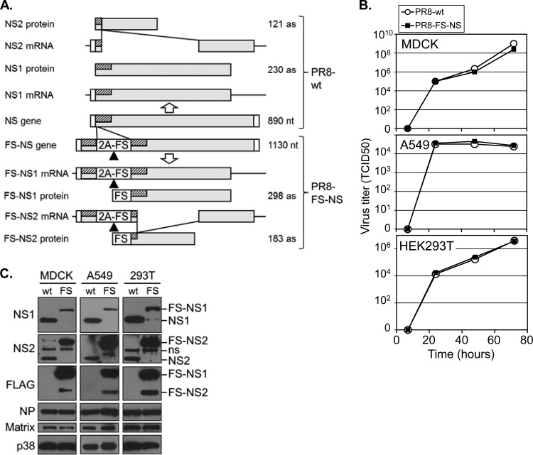
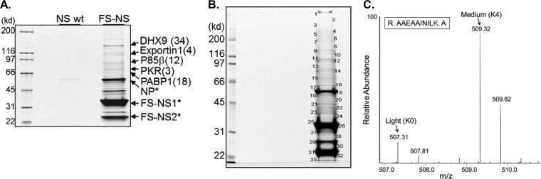
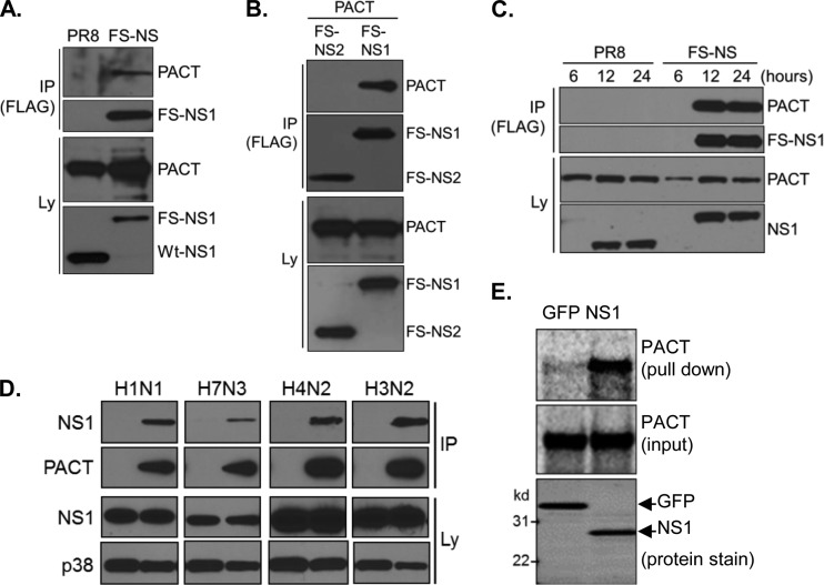
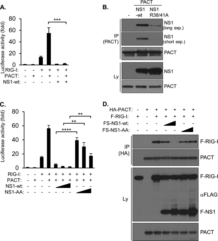
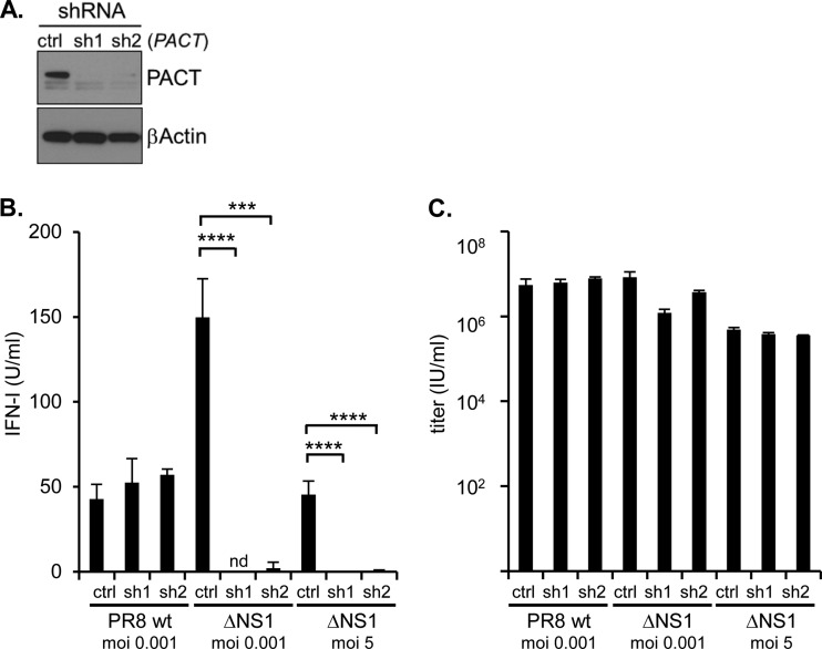
Influenza A virus (IAV) replication depends on the interaction of virus proteins with host factors. The viral nonstructural protein 1 (NS1) is essential in this process by targeting diverse cellular functions, including mRNA splicing and translation, cell survival, and immune defense, in particular the type I interferon (IFN-I) response. In order to identify host proteins targeted by NS1, we established a replication-competent recombinant IAV that expresses epitope-tagged forms of NS1 and NS2, which are encoded by the same gene segment, allowing purification of NS proteins during natural cell infection and analysis of interacting proteins by quantitative mass spectrometry. We identified known NS1- and NS2-interacting proteins but also uncharacterized proteins, including PACT, an important cofactor for the IFN-I response triggered by the viral RNA-sensor RIG-I. We show here that NS1 binds PACT during virus replication and blocks PACT/RIG-I-mediated activation of IFN-I, which represents a critical event for the host defense. Protein interaction and interference with IFN-I activation depended on the functional integrity of the highly conserved RNA binding domain of NS1. A mutant virus with deletion of NS1 induced high levels of IFN-I in control cells, as expected; in contrast, shRNA-mediated knockdown of PACT compromised IFN-I activation by the mutant virus, but not wild-type virus, a finding consistent with the interpretation that PACT (i) is essential for IAV recognition and (ii) is functionally compromised by NS1. Together, our data describe a novel approach to identify virus-host protein interactions and demonstrate that NS1 interferes with PACT, whose function is critical for robust IFN-I production.

Importance: Influenza A virus (IAV) is an important human pathogen that is responsible for annual epidemics and occasional devastating pandemics. Viral replication and pathogenicity depends on the interference of viral factors with components of the host defense system, particularly the type I interferon (IFN-I) response. The viral NS1 protein is known to counteract virus recognition and IFN-I production, but the molecular mechanism is only partially defined. We used a novel proteomic approach to identify host proteins that are bound by NS1 during virus replication and identified the protein PACT, which had previously been shown to be involved in virus-mediated IFN-I activation. We find that NS1 prevents PACT from interacting with an essential component of the virus recognition pathway, RIG-I, thereby disabling efficient IFN-I production. These observations provide an important piece of information on how IAV efficiently counteracts the host immune defense.


Figures

FIG 1
FIG 1
Generation of a recombinant PR8 virus with epitope-tagged NS1- and NS2 (PR8-NS-FS). (A) IAV NS gene arrangement and structure of recombinant PR8-NS-FS virus through the duplication of 90 bp of the 5′ gene segment (hatched), followed by the 2A peptide of the porcine teschovirus-1 (P2A) and a tandem triple-FLAG-StrepOne epitope tag (FS). Different expected mRNA and protein products for PR8-wt (upper part) and PR8-NS-FS (lower part) are shown. Triangle, 2A-encoded “cleavage” site. (B) Replication of PR8-wt and PR8 NS-FS on MDCK, A549, and HEK293T cells was determined by infection at an MOI of 0.001 (MDCK and A549 cells) or at an MOI of 0.01 (HEK293T cells) and determination of the virus titer from the supernatants at the indicated time points. (C) Immunoblot analysis of MDCK, A549, and HEK293T cells, infected with PR8-wt (wt) and PR8-NS-FS (FS) at an MOI of 1 for 24 h. Blots were analyzed with antibodies against proteins indicated (left), and the protein identity is indicated (right).
FIG 2
FIG 2
Purification of NS1/2 from IAV-infected cells and identification of associated proteins by MS. (A) SYPRO Ruby-stained SDS-PAGE of samples derived from A549 cells that were infected with PR8-wt and PR8-FS-NS for 24 h at an MOI of 0.5, followed by TAP. (B) SYPRO Ruby-stained SDS-PAGE of purified protein samples from SILAC-labeled HEK293T cells. HEK293T cells were labeled with Lys0/Arg0 (light) and Lys4/Arg6 (medium) and infected with PR8-wt (light) or PR8-NS-FS (medium), followed by protein purification and SDS-PAGE. (C) Bands subjected to LC-MS/MS analysis are indicated. Mass spectrum of a peptide representing PACT, whose differentially labeled forms (Light from PR8-wt-infected cells and medium from PR8-NS-FS-infected cells) can be identified by their specific mass difference of 4 Da (= 2 m/z of double-charged peptide, for lysine). Arrows indicate the C12 monoisotopic peaks of each peptide.
FIG 3
FIG 3
NS1 interacts with PACT during virus replication and in vitro. (A) HEK293T cells were infected with PR8-wt or PR8 NS-FS for 24 h, followed by immunoprecipitation (IP) and immunoblotting (IB) with antibodies against PACT and NS1 (Ly, total lysate). (B) HEK293T cells were transfected with PACT along with FS-tagged NS1 or NS2 for 48 h, followed by IP/IB analysis using antibodies against FLAG (for IP) and antibodies against FLAG (detecting FS-NS1 and FS-NS2) and PACT (for IB). (C) HEK293T cells were infected with PR8-wt or PR8 NS-FS for 6, 12, or 24 h, followed by IP and IB with antibodies against FLAG (for IP) and PACT and NS1 (for IB). (D) HEK293T cells were transfected with control plasmid or HA-tagged PACT, followed by infection with indicated IAV strains for 24 h and IP/IB analysis using antibodies against HA (for IP) and PACT, NS1, and p38 (for IB). H1N1, PR8; H7N3, A/laughing gull/Delaware bay/42/2006; H4N2, A/quail/California/D113023808/2012; H3N2 (A/Perth/16/2009). (E) Pulldown experiment of in vitro-translated PACT using recombinant, bead-immobilized GFP or NS1. A pull-down sample and 10% of input sample were resolved by SDS-PAGE and analyzed by using a phosphorimager or SYPRO Ruby protein staining, respectively.
FIG 4
FIG 4
NS1 prevents interaction between PACT and RIG-I and blocks PACT/RIG-I-mediated IFN-β activation. (A) IFN-β reporter assay. HEK293T cells were transfected with an IFN-β promoter luciferase vector, either alone (control) or along with expression vectors for RIG-I, PACT, and NS1, as indicated, followed by analysis of luciferase activity after 48 h. Values are presented as the fold changes over the control. Error bars represent the standard deviations of three transfection replicates. ***, P < 0.001. (B) HEK293T cells transfected with PACT, along with FS-NS1 or FS-NS1 (R38A/K41A) (NS1-AA), for 48 h, followed by IP/IB analysis with antibodies against PACT (for IP) and NS1 and PACT (for IB). Long and short exposures of films are shown, as indicated. (C) IFN-β reporter assay. HEK293T cells were transfected with an IFN-β promoter luciferase vector, either alone (control) or along with expression vectors for RIG-I, PACT, NS1, and NS1-AA as indicated, followed by analysis of luciferase activity after 48 h. Values are presented as the fold changes over the control. Error bars represent the standard deviations of three transfection replicates. **, P < 0.01; ****, P < 0.0001. (D) HEK293T cells were transfected with expression vectors for the indicated proteins for 48 h and analyzed by IP/IB with antibodies against HA (for IP) and antibodies against RIG-I, PACT, and FLAG (recognizing FLAG-RIG-I and FS-NS1) (for IB).
FIG 5
FIG 5
PACT is required for efficient IFN-I production upon IVA infection in the absence of NS1. (A) Stable knockdown of PACT in HEK293T cells by two different, retrovirally delivered shRNAs against PACT mRNA (sh1, sh2) and control shRNA (ctrl), analyzed by IB with antibodies against PACT and β-actin. (B and C) The HEK293T cells described in panel A were infected with PR8-wt and PR8 ΔNS1 at an MOI of 0.001 for 72 h or with PR8 ΔNS1 at an MOI of 5 for 24 h as indicated. The IFN-I activity (B) and virus titer (C) were determined by IFN bioassay and limiting dilution, after 72 h (MOI of 0.001) and 24 h (MOI of 5), respectively. nd, not detectable; ***, P < 0.001; ****, P < 0.0001.

References

    1. Hale BG, Randall RE, Ortin J, Jackson D. 2008. The multifunctional NS1 protein of influenza A viruses. The J. Gen. Virol. 89:2359–2376. 10.1099/vir.0.2008/004606-0 - DOI - PubMed
    1. Egorov A, Brandt S, Sereinig S, Romanova J, Ferko B, Katinger D, Grassauer A, Alexandrova G, Katinger H, Muster T. 1998. Transfectant influenza A viruses with long deletions in the NS1 protein grow efficiently in Vero cells. J. Virol. 72:6437–6441 - PMC - PubMed
    1. Kochs G, Koerner I, Thiel L, Kothlow S, Kaspers B, Ruggli N, Summerfield A, Pavlovic J, Stech J, Staeheli P. 2007. Properties of H7N7 influenza A virus strain SC35M lacking interferon antagonist NS1 in mice and chickens. J. Gen. Virol. 88:1403–1409. 10.1099/vir.0.82764-0 - DOI - PubMed
    1. Garcia-Sastre A, Egorov A, Matassov D, Brandt S, Levy DE, Durbin JE, Palese P, Muster T. 1998. Influenza A virus lacking the NS1 gene replicates in interferon-deficient systems. Virology 252:324–330. 10.1006/viro.1998.9508 - DOI - PubMed
    1. Lu Y, Wambach M, Katze MG, Krug RM. 1995. Binding of the influenza virus NS1 protein to double-stranded RNA inhibits the activation of the protein kinase that phosphorylates the elF-2 translation initiation factor. Virology 214:222–228. 10.1006/viro.1995.9937 - DOI - PubMed

Publication types

MeSH terms