. 2014 Aug;88(16):9100-10.
doi: 10.1128/JVI.01129-14. Epub 2014 Jun 4.

Extraribosomal l13a is a specific innate immune factor for antiviral defense

Affiliations

Extraribosomal l13a is a specific innate immune factor for antiviral defense

Barsanjit Mazumder et al. J Virol. 2014 Aug.

Abstract

We report a novel extraribosomal innate immune function of mammalian ribosomal protein L13a, whereby it acts as an antiviral agent. We found that L13a is released from the 60S ribosomal subunit in response to infection by respiratory syncytial virus (RSV), an RNA virus of the Pneumovirus genus and a serious lung pathogen. Unexpectedly, the growth of RSV was highly enhanced in L13a-knocked-down cells of various lineages as well as in L13a knockout macrophages from mice. In all L13a-deficient cells tested, translation of RSV matrix (M) protein was specifically stimulated, as judged by a greater abundance of M protein and greater association of the M mRNA with polyribosomes, while general translation was unaffected. In silico RNA folding analysis and translational reporter assays revealed a putative hairpin in the 3'untranslated region (UTR) of M mRNA with significant structural similarity to the cellular GAIT (gamma-activated inhibitor of translation) RNA hairpin, previously shown to be responsible for assembling a large, L13a-containing ribonucleoprotein complex that promoted translational silencing in gamma interferon (IFN-γ)-activated myeloid cells. However, RNA-protein interaction studies revealed that this complex, which we named VAIT (respiratory syncytial virus-activated inhibitor of translation) is functionally different from the GAIT complex. VAIT is the first report of an extraribosomal L13a-mediated, IFN-γ-independent innate antiviral complex triggered in response to virus infection. We provide a model in which the VAIT complex strongly hinders RSV replication by inhibiting the translation of the rate-limiting viral M protein, which is a new paradigm in antiviral defense.

Importance: The innate immune mechanisms of host cells are diverse in nature and act as a broad-spectrum cellular defense against viruses. Here, we report a novel innate immune mechanism functioning against respiratory syncytial virus (RSV), in which the cellular ribosomal protein L13a is released from the large ribosomal subunit soon after infection and inhibits the translation of a specific viral mRNA, namely, that of the matrix protein M. Regarding its mechanism, we show that the recognition of a specific secondary structure in the 3' untranslated region of the M mRNA leads to translational arrest of the mRNA. We also show that the level of M protein in the infected cell is rate limiting for viral morphogenesis, providing a rationale for L13a to target the M mRNA for suppression of RSV growth. Translational silencing of a viral mRNA by a deployed ribosomal protein is a new paradigm in innate immunity.


Figures

FIG 1
FIG 1
L13 deficiency leads to better virus growth. (A, left) Five independent A549 cell clones with stably integrated anti-L13a shRNA (1 to 5) or control nonspecific shRNA (denoted C) were infected with RSV, and the total proteins of infected cell monolayers at 18 hpi were analyzed for L13a (and control actin) by immunoblotting. Clones 1 to 4 used shRNA against the coding sequence; however, in clone 5 the shRNA was against the 3′UTR, and thus, L13a could be restored by transient transfection with FLAG-L13a plasmid (1 μg DNA per 5 × 10⁵ cells), and virus was added 24 h after transfection. In parallel wells, virus liberated in cell-free medium at 72 hpi was quantified by standard serial dilution and plaque assay on HEp-2 cells. Note the clonal variation of L13 silencing and the inverse correlation between L13a levels and viral titer. (A, right) As described above for the left panel, except that macrophages from two L13a KO mice (L13aflox/floxLysMCre+) (KO-1 and KO-2) and two control mice (L13aflox/flox) (C-1 and C-2) were harvested and used in RSV infection. (B) Infectious virus, liberated in the medium at different times following infection of cultured cells, was plaque assayed on HEp-2 cells. Numbers from the two KO macrophage lines and the two controls were averaged and plotted with error bars and the P value as shown. (C) RSV infection foci formed on control and L13a KO macrophages. We used a low MOI (∼0.5) for better recognition of the foci in these cells. Top, photomicrographs of the infected macrophages. Note that macrophages are relatively round and loosely attached to the plastic surface; thus, in contrast to the large syncytia that are characteristically formed in RSV-infected epithelial cell monolayers, the infected macrophages produce clumps or foci. The average number (with standard error) of recognizable foci in multiple fields of the same area is written below each image and is similar in control (18 ± 4) and KO (19 ± 3) cells. However, note the larger size of the foci in the KO, consistent with reinfection of a greater number of neighboring cells by the larger number of released progeny virus (A and B). Bottom, the diameters of 200 foci were measured in control (dark line) and L13a-KO (gray line) fields, and the percentage of each size category was plotted for those that were above 1%.
FIG 2
FIG 2
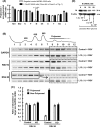
L13a deficiency rescues the polysomal association of viral M mRNA. (A) Polysomal association in L13 knockdown A549 cells. Ribosomal fractions isolated from RSV-infected L13a-silenced A549 clone 4 and control cells (Fig. 1A) were subjected to 5 to 25% sucrose gradient centrifugation as described in Materials and Methods (12). Total RNA from each fraction was purified and used as the template for real-time RT-PCR with the gene-specific primers described in Materials and Methods. For each gene, the ratio of polysomal to nonpolysomal mRNA amount was calculated and plotted. Note the uniquely low polysomal abundance of the M mRNA in the control cell and its restoration in L13a-deficient cells, the highly significant difference being indicated by the low P value. Average results from three experiments are presented with standard error bars. (B) Polysomal association profile of selected mRNA in L13a knockout and control mouse macrophages. Bone marrow-derived macrophages were infected with RSV, and polysome fractionation and RT-PCR were performed as described for panel A. Representative agarose gel profiles of the ethidium bromide-stained PCR products are shown. A plot of the A254 values of the fractions (1 through 12) is shown at the top. Note the shift of a large pool of M mRNA from the nonpolysomal (fractions 1 to 5; solid line box) to the polysomal (fractions 8 to 12; dashed line box) region upon loss of L13a, whereas the distributions of two control mRNAs, namely, viral N and cellular GAPDH, were not affected. (C) Quantification of the results shown in panel B. Ratios of target mRNA/GAPDH mRNA in polysomal and nonpolysomal fractions were determined by measuring the intensities of the corresponding bands for two viral mRNAs, L13-regulated M and control N. (D) RNA interference (RNAi)-mediated knockdown of M protein severely inhibits RSV growth. RSV infection was carried out in A549 cells as described for Fig. 1A, except that the virus was added 6 h after transfection with increasing concentrations (0 to 50 nM) of anti-M siRNA1 (see Materials and Methods). Silencing of M was confirmed by immunoblotting, while profilin (control) was unaffected. The virus liberated in the medium at 48 h postinfection was plaque assayed on an HEp-2 cell layer as described for Fig. 1A. Use of anti-M siRNA2 generated essentially identical results (not shown).
FIG 3
FIG 3
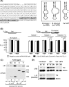
Translational repression by L13a requires cis-acting sequences of the M mRNA. (A) Two regions of RSV A serotype M mRNA (left) with potential hairpin structures predicted by Mfold (right) (12, 27). The start and the stop codons are in lowercase; the two hairpin sequences are underlined. (B) The M hairpin region inhibits translation in cis in RSV-infected cells in an L13a-dependent manner. The 243-nt-long sequence near the 3′-end (A) was cloned downstream of the luciferase reporter construct as shown, the DNA was transfected into A549 cells (or A549 L13a knockdown clone 4), and Luc activity was measured with or without RSV infection at 24 h postinfection. Control cells (left) were transfected with Luc plasmid without the M sequence and treated similarly. Significant differences are indicated by the P values. (C) Recombinantly overexpressed M protein alone stimulates RSV growth. A549 cells were transiently transfected with M expression clones with (M-U) or without (M) 3′UTR or with N and P expression clones and also infected with RSV. Total M protein was detected by immunoblotting with a polyclonal antibody against full-length M, and all recombinant proteins, which were FLAG tagged, were detected by monoclonal FLAG antibody. All protein bands are marked with their molecular mass values: 30 kDa (M), 38 kDa (P), and 42 kDa (N). The first lane (—) represents control, untransfected but RSV-infected A549 cells. (D) L13a is rapidly released from ribosomes in RSV-infected epithelial cells, as detected by immunoblotting; control L11 protein was not released. Infections with VSV and hMPV, carried out similarly, did not promote L13a release, even at 24 hpi.
FIG 4
FIG 4
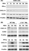
The RSV (VAIT) mechanism functionally differs from that of GAIT. A549 cells (A) or primary macrophages (Mϕ) (B) were either infected with RSV (at an MOI of 3) or treated with IFN-γ (500 units/ml) as shown, and at various times thereafter, total cell lysates were prepared and subjected to immunoblotting to detect the indicated proteins. Note the induction of L13-regulated CXCL13 and CCR3 in IFN-γ-treated macrophages but no such induction upon RSV infection.
FIG 5
FIG 5
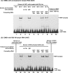
Specific RNP complexes formed by GAIT (A) and VAIT (B) RNA. EMSA with synthetic biotin-labeled RNA was performed as described in Materials and Methods, using extracts of either RSV-infected A549 cells (lanes 9, 10, and 12 to 18) or U937 cells (lanes 1 to 8 and 19) grown in the presence of IFN-γ (500 units/ml) for 8 h or 24 h, as shown. Where indicated, the extract was preincubated with either rabbit polyclonal L13a antibody (αL13a) (lanes 3 and 13) or control nonimmune immunoglobulin (IgG) (lanes 4 and 14). For competition, the same GAIT or VAIT RNAs, but not labeled with biotin, were used in either 10-fold or 100-fold excess as indicated. In both panels, the positions of the mobility shifted (retarded) RNA band (RNP complex) and the free RNA are marked.
FIG 6
FIG 6
Model for regulation of RSV growth by L13a. An unknown but potentially novel signaling mechanism promotes rapid release of L13a from RSV-infected cell ribosomes, and this extraribosomal L13a then silences viral M translation by recruiting an RNP complex at the VAIT hairpin in the 3′UTR (see the text for details). We present this cellular response as an innate immune mechanism that moderates virus growth and possibly the associated pathology of the host.

Similar articles

Cited by

References

    1. Collins PL, Melero JA. 2011. Progress in understanding and controlling respiratory syncytial virus: still crazy after all these years. Virus Res. 162:80–99. 10.1016/j.virusres.2011.09.020 - DOI - PMC - PubMed
    1. Sommer C, Resch B, Simões EA. 2011. Risk factors for severe respiratory syncytial virus lower respiratory tract infection. Open Microbiol. J. 5:144–154. 10.2174/1874285801105010144 - DOI - PMC - PubMed
    1. Graham BS. 2011. Biological challenges and technological opportunities for respiratory syncytial virus vaccine development. Immunol. Rev. 239:149–166. 10.1111/j.1600-065X.2010.00972.x - DOI - PMC - PubMed
    1. Barik S. 2013. Respiratory syncytial virus mechanisms to interfere with type 1 interferons. Curr. Top. Microbiol. Immunol. 372:173–191. 10.1007/978-3-642-38919-1_9 - DOI - PubMed
    1. Kumar H, Kawai T, Akira S. 2011. Pathogen recognition by the innate immune system. Int. Rev. Immunol. 30:16–34. 10.3109/08830185.2010.529976 - DOI - PubMed

Publication types

MeSH terms

LinkOut - more resources