. 2014 Aug;88(16):9245-59.
doi: 10.1128/JVI.00841-14. Epub 2014 Jun 4.

The gammaherpesviruses Kaposi's sarcoma-associated herpesvirus and murine gammaherpesvirus 68 modulate the Toll-like receptor-induced proinflammatory cytokine response

Affiliations

The gammaherpesviruses Kaposi's sarcoma-associated herpesvirus and murine gammaherpesvirus 68 modulate the Toll-like receptor-induced proinflammatory cytokine response

Kendra A Bussey et al. J Virol. 2014 Aug.

Abstract

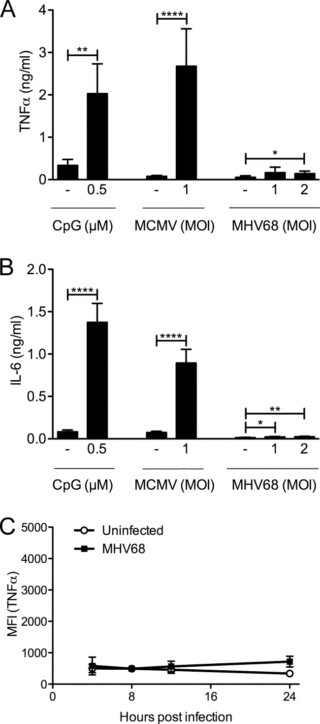
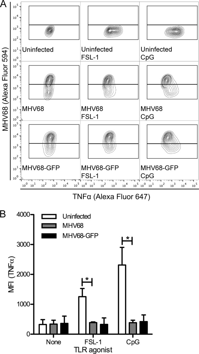
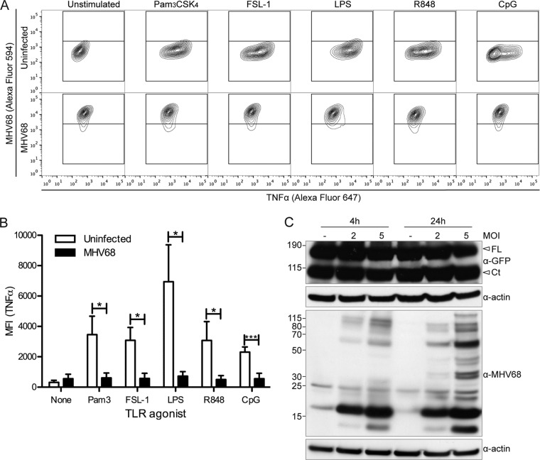
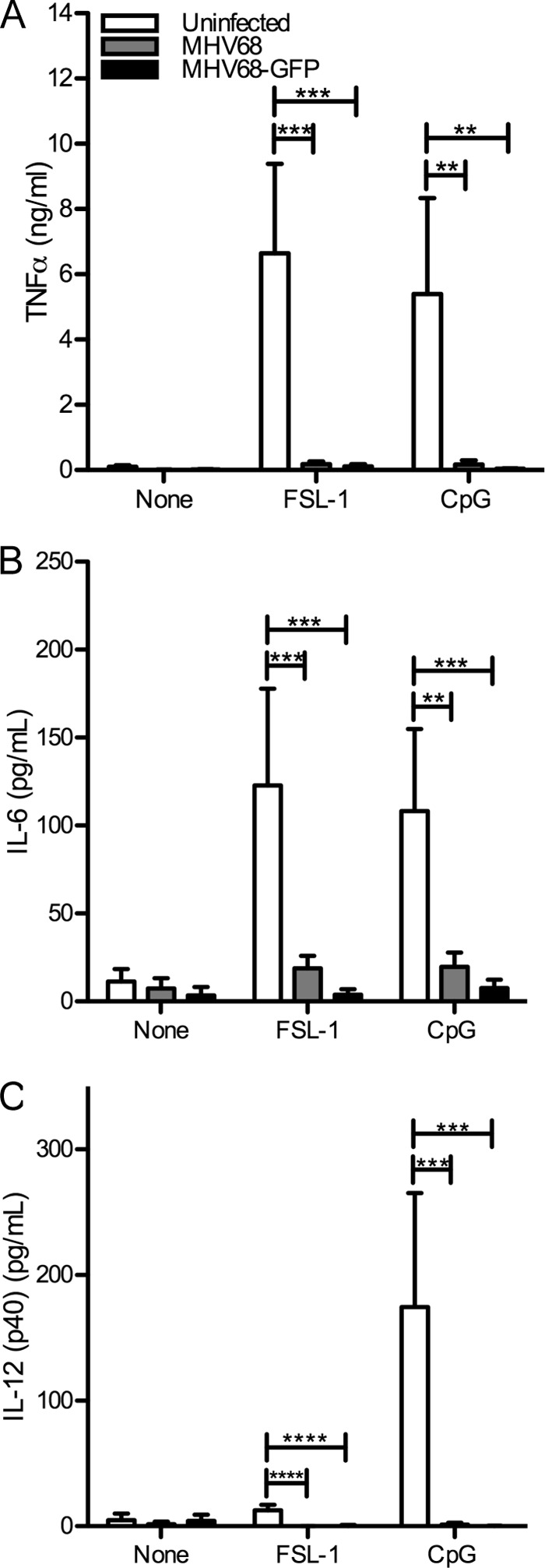
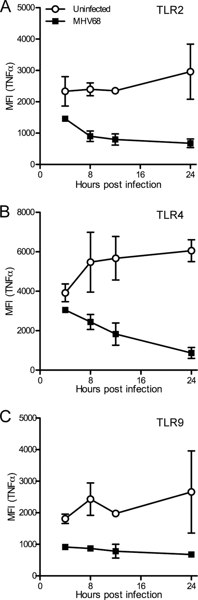
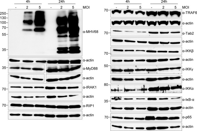
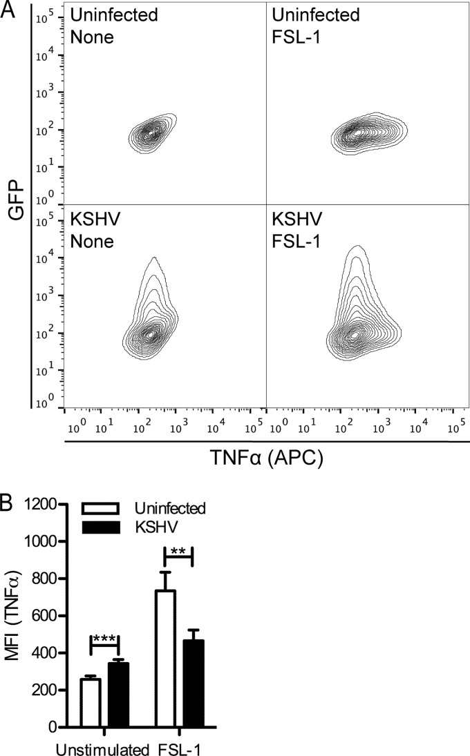
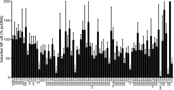
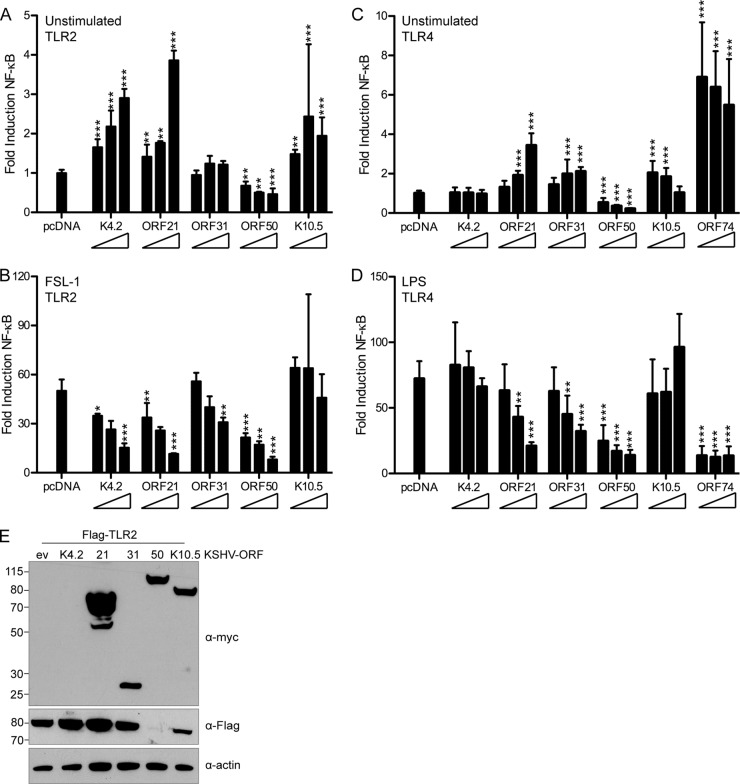
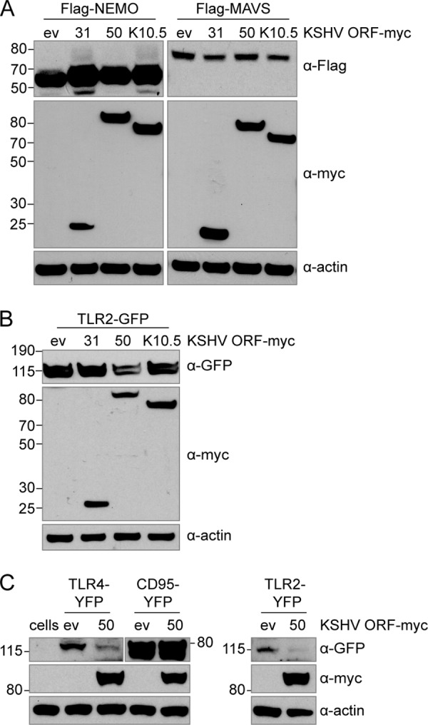
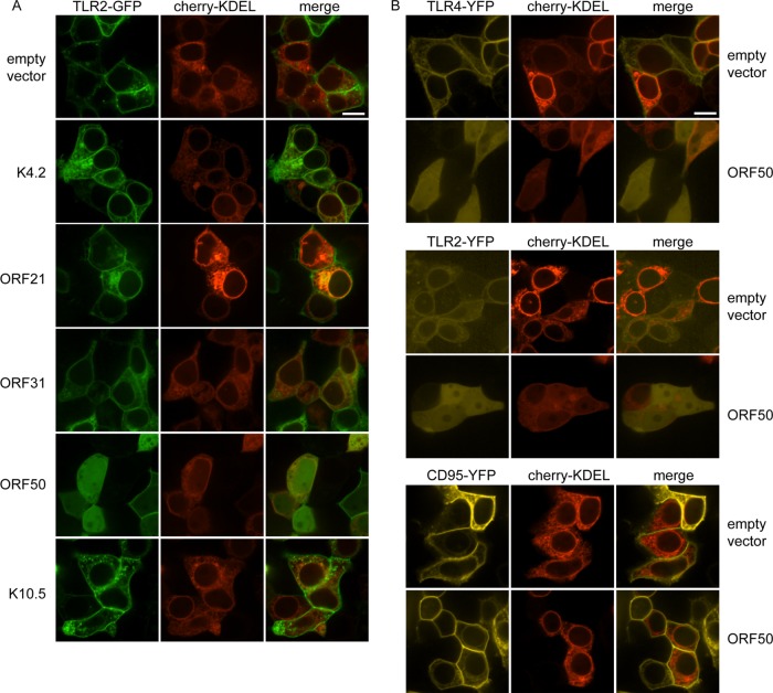
The human pathogen Kaposi's sarcoma-associated herpesvirus (KSHV), the etiological agent of Kaposi's sarcoma, primary effusion lymphoma, and multicentric Castleman's disease, establishes lifelong latency upon infection. Murine gammaherpesvirus 68 (MHV68) is a well-established model for KSHV. Toll-like receptors (TLRs) play a crucial role for the innate immune response to pathogens. Although KSHV and MHV68 are detected by TLRs, studies suggest they modulate TLR4 and TLR9 signaling, respectively. In this study, we show that in bone marrow-derived macrophages (BMDMs), MHV68 did not induce a detectable proinflammatory cytokine response. Furthermore, MHV68 abrogated the response to TLR2, -4, -7, and -9 agonists in BMDMs. Similarly to observations with MHV68, infection with KSHV efficiently inhibited TLR2 signaling in THP-1 monocytes. Using a KSHV open reading frame (ORF) library, we found that K4.2, ORF21, ORF31, and the replication and transcription activator protein (RTA)/ORF50 inhibited TLR2-dependent nuclear factor kappa B (NF-κB) activation in HEK293 TLR2-yellow fluorescent protein (YFP)- and Flag-TLR2-transfected HEK293T cells. Of the identified ORFs, RTA/ORF50 strongly downregulated TLR2 and TLR4 signaling by reducing TLR2 and TLR4 protein expression. Confocal microscopy revealed that TLR2 and TLR4 were no longer localized to the plasma membrane in cells expressing RTA/ORF50. In this study, we have shown that the gammaherpesviruses MHV68 and KSHV efficiently downmodulate TLR signaling in macrophages and have identified a novel function of RTA/ORF50 in modulation of the innate immune response.

Importance: The Toll-like receptors (TLRs) are an important class of pattern recognition receptors of the innate immune system. They induce a potent proinflammatory cytokine response upon detection of a variety of pathogens. In this study, we found that the gammaherpesviruses murine gammaherpesvirus 68 (MHV68) and Kaposi's sarcoma-associated herpesvirus (KSHV) efficiently inhibit the TLR-mediated innate immune response. We further identified the KSHV-encoded replication and transcription activator protein (RTA) as a novel modulator of TLR signaling. Our data suggest that the gammaherpesviruses MHV68 and KSHV prevent activation of the innate immune response by targeting TLR signaling.


Figures

FIG 1
FIG 1
The proinflammatory cytokine response of BMDMs to MHV68 is very weak. Primary BMDMs were stimulated with CpG B or infected with MCMV-GFP or MHV68 for 16 h. TNF-α (A) or IL-6 (B) production was detected by ELISA. Results are shown as means + standard deviations (SD) from duplicates from 2 (CpG B, A) or 3 independent experiments. Immortalized BMDMs were infected for 4 to 24 h with MHV68-GFP (MOI of 5) (C). Four hours prior to harvesting, cells were incubated with brefeldin A to prevent TNF-α secretion and then fixed and processed for intracellular TNF-α staining. Samples were analyzed by flow cytometry. The means ± SD of mean fluorescence intensity (MFI) for the anti-TNF-α signal from 2 independent experiments are shown. *, P < 0.05; **, P < 0.01; ****, P < 0.0001.
FIG 2
FIG 2
MHV68-infected primary BMDMs do not respond to TLR stimulation. At 24 h postinfection, MHV68- or MHV68-GFP-infected (MOI of 2) or uninfected primary BMDMs were stimulated for 4 h with TLR agonists (TLR2, FSL-1; TLR9, CpG B) in the presence of brefeldin A to prevent TNF-α secretion and then processed for intracellular MHV68 and TNF-α staining and analyzed by flow cytometry. (A) Representative contour plots. (B) The averages + SD of the mean fluorescence intensity of the anti-TNF-α channel gated on MHV68-negative (uninfected) or MHV68-positive (MHV68 or MHV68-GFP) populations from 2 independent experiments are shown. *, P < 0.05.
FIG 3
FIG 3
MHV68-infected immortalized BMDMs do not respond to TLR stimulation. (A, B) At 24 h postinfection, MHV68-GFP-infected (MOI of 5) or uninfected immortalized BMDMs were stimulated for 4 h with TLR agonists (TLR2, Pam3CSK4, FSL-1; TLR4, LPS; TLR7, R848; TLR9, CpG B) in the presence of brefeldin A to prevent TNF-α secretion and then processed for intracellular TNF-α staining and analyzed by flow cytometry. (A) Representative contour plots (uninfected, upper row; MHV68-GFP infected, lower row). (B) The averages + SD of the mean fluorescence intensity of the anti-TNF-α channel gated on MHV68-negative (uninfected) or MHV68-positive (MHV68 or MHV68-GFP) populations from 3 independent experiments are shown. *, P < 0.05; ***, P < 0.001. (C) Immortalized BMDMs expressing TLR9-GFP were infected with MHV68 at the indicated MOIs or left uninfected, and then TLR9 levels at 4 h and 24 h postinfection were determined by immunoblotting with an anti-GFP antibody (FL, full-length TLR9-GFP; Ct, C-terminal cleavage fragment of TLR9-GFP). Immunoblots for MHV68 and actin are included as controls.
FIG 4
FIG 4
MHV68-infected BMDMs do not produce proinflammatory cytokines in response to TLR stimulation. At 8 h postinfection, MHV68- or MHV68-GFP-infected (MOI of 5) or uninfected immortalized BMDMs were stimulated with TLR agonists (TLR2, FSL-1; TLR9, CpG B) for 16 h, and then TNF-α (A), IL-6 (B), and IL-12 (p40) (C) levels were determined by ELISA. The averages + SD from triplicate samples from 2 (A, C) or 4 (B) independent infection experiments are shown. **, P < 0.01; ***, P < 0.001; ****, P < 0.0001.
FIG 5
FIG 5
MHV68 inhibits TLR-induced TNF-α production in BMDMs early after infection. Immortalized BMDMs were infected for 4 to 24 h with MHV68-GFP (MOI of 5). Four hours prior to harvesting, cells were incubated with TLR agonists for TLR2 (FSL-1; A), TLR4 (LPS; B), or TLR9 (CpG B; C) in the presence of brefeldin A to prevent TNF-α secretion and then fixed and processed for intracellular TNF-α staining. Samples were analyzed by flow cytometry. The means ± SD of mean fluorescence intensity (MFI) for the anti-TNF-α signal from 2 independent experiments are shown. For reference, the MFI for unstimulated samples was at all times less than 1,000 (see Fig. 1C). For each time course, P < 0.0001.
FIG 6
FIG 6
Steady-state expression levels of proteins involved in TLR signaling are not affected in MHV68-infected cells. Immortalized BMDMs were infected with MHV68 at an MOI of 2 or 5 or left uninfected and then lysed at 4 and 24 h with RIPA buffer. Equal volumes of lysates were separated by SDS-PAGE or Bis-Tris PAGE and transferred to nitrocellulose membranes, and then protein levels for the indicated proteins were determined by immunoblotting with the indicated antibodies. Separate gels were used for each antibody, and actin blots are included as controls. Results are representative of 3 independent experiments.
FIG 7
FIG 7
KSHV inhibits the TLR2-induced TNF-α response in THP-1 monocytes. THP-1 cells were treated with Polybrene or infected with GFP-expressing KSHVLyt (MOI of 0.5) in the presence of Polybrene. Twenty-four hours postinfection, cells were stimulated or not with FSL-1 in the presence of brefeldin A and then processed for TNF-α intracellular cytokine staining. (A) Representative contour plots. (B) The averages + SD of mean fluorescence intensity (MFI) of duplicates from 2 independent experiments are shown. For KSHVLyt-infected samples, MFI was determined for the GFP-positive population. **, P < 0.01; ***, P < 0.001.
FIG 8
FIG 8
Screen for KSHV ORFs that may inhibit the TLR2-induced NF-κB response. HEK293 TLR2-YFP cells were transfected with pNF-κB-Luc (firefly), pTK-RL (renilla), and the indicated KSHV ORF-Myc or empty pcDNA vector. At 24 h posttransfection, cells were unstimulated or stimulated with the TLR2 agonist FSL-1 overnight and then lysed in passive lysis buffer for measurement of luciferase activity. Fold induction of NF-κB is determined as follows: firefly normalized to renilla and then stimulated divided by unstimulated, scaled to pcDNA empty vector (100%). Data shown are the averages from 4 replicates total from 3 independent experiments. Log-transformed results were analyzed by one-way ANOVA (P < 0.0001), and select empty vector-ORF pairs were analyzed by two-tailed unpaired t test. ***, P < 0.001; ****, P < 0.0001.
FIG 9
FIG 9
ORF21 and ORF50 strongly inhibit TLR2 and TLR4 signaling. (A, B) HEK293T cells were cotransfected with pNF-κB-Luc, pTK-RL, Flag-TLR2, and empty pcDNA vector or increasing concentrations (1×, 2×, or 4×) of the indicated KSHV ORF-Myc. At 18 h posttransfection, cells were unstimulated or stimulated for 6 h with the TLR2 agonist FSL-1 and then lysed in passive lysis buffer. (C, D) HEK293 TLR4-MD2-CD14 cells were cotransfected with pNF-κB-Luc, pTK-RL, and empty pcDNA vector or increasing concentrations (1×, 2×, or 4×) of the indicated KSHV ORF-Myc. At 18 h posttransfection, cells were unstimulated or stimulated for 9 h with the TLR4 agonist LPS and then lysed in passive lysis buffer for measurement of luciferase activity. (A, C) NF-κB response in unstimulated cells, normalized to renilla and scaled to empty vector. (B, D) Fold induction of NF-κB in response to FSL-1 (B) or LPS (D), determined as follows: firefly normalized to renilla and then stimulated divided by unstimulated. Data shown are averages with standard deviations from 3 or 4 replicates total in two independent experiments (A, B) or duplicates from each of 3 independent experiments (C, D). Transformed results were analyzed by one-way ANOVA followed by Dunnett's multiple comparison test. *, P < 0.05; **, P < 0.01; ***, P < 0.001. (E) Flag and Myc immunoblotting was used to verify Flag-TLR2 and ORF-Myc expression levels in whole-cell RIPA lysates. Actin was used as a loading control.
FIG 10
FIG 10
ORF50 specifically affects protein levels of TLR2 and TLR4. HEK293T cells were cotransfected with Myc-tagged KSHV ORFs (ORF31, ORF50, or K10.5) and empty pcDNA vector and Flag-tagged MAVS, Flag-tagged NEMO (A), or TLR2-GFP (B). HEK293T cells were cotransfected with ORF50 or empty pcDNA vector and TLR4-YFP, CD95-YFP, or TLR2-YFP. (C) Nontransfected cells were used as a negative control. Protein levels at 24 h posttransfection in whole-cell lysates were determined by immunoblotting using anti-Flag (A), anti-GFP ab290 (B), or anti-GFP D5.1 (C) and anti-Myc and anti-actin (A, B, C) antibodies.
FIG 11
FIG 11
In ORF50-expressing cells, TLR2 and TLR4 no longer localize to the plasma membrane. HEK293T cells were cotransfected with the indicated plasmids and images were obtained 24 h posttransfection in live cells by confocal microscopy. Cherry-KDEL (red) was used as a marker for cotransfection and the endoplasmic reticulum and included in all transfections. Cells were cotransfected with TLR2-GFP and either empty pcDNA vector, K4.2, ORF21, ORF31, ORF50, or K10.5 (A) or with combinations of TLR4-YFP, TLR2-YFP, or CD95-YFP and empty pcDNA vector or ORF50 (B). Scale bar, 10 μm.

References

    1. Chang Y, Cesarman E, Pessin MS, Lee F, Culpepper J, Knowles DM, Moore PS. 1994. Identification of herpesvirus-like DNA sequences in AIDS-associated Kaposi's sarcoma. Science 266:1865–1869. 10.1126/science.7997879 - DOI - PubMed
    1. Dittmer DP, Damania B. 2013. Kaposi sarcoma associated herpesvirus pathogenesis (KSHV)—an update. Curr. Opin. Virol. 3:238–244. 10.1016/j.coviro.2013.05.012 - DOI - PMC - PubMed
    1. Blaskovic D, Stancekova M, Svobodova J, Mistrikova J. 1980. Isolation of five strains of herpesviruses from two species of free living small rodents. Acta Virol. 24:468. - PubMed
    1. Barton E, Mandal P, Speck SH. 2011. Pathogenesis and host control of gammaherpesviruses: lessons from the mouse. Annu. Rev. Immunol. 29:351–397. 10.1146/annurev-immunol-072710-081639 - DOI - PubMed
    1. Knipe DM, Howley PM. 2013. Fields virology, 6th ed, vol II Wolters Kluwer, Philadelphia, PA

Publication types

MeSH terms

LinkOut - more resources