. 2014 Aug;88(16):9111-28.
doi: 10.1128/JVI.01502-14. Epub 2014 Jun 4.

Maturation and vesicle-mediated egress of primate gammaherpesvirus rhesus monkey rhadinovirus require inner tegument protein ORF52

Affiliations

Maturation and vesicle-mediated egress of primate gammaherpesvirus rhesus monkey rhadinovirus require inner tegument protein ORF52

Melissa S Anderson et al. J Virol. 2014 Aug.

Abstract

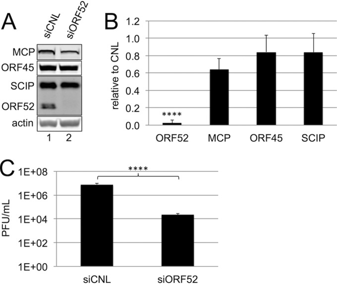
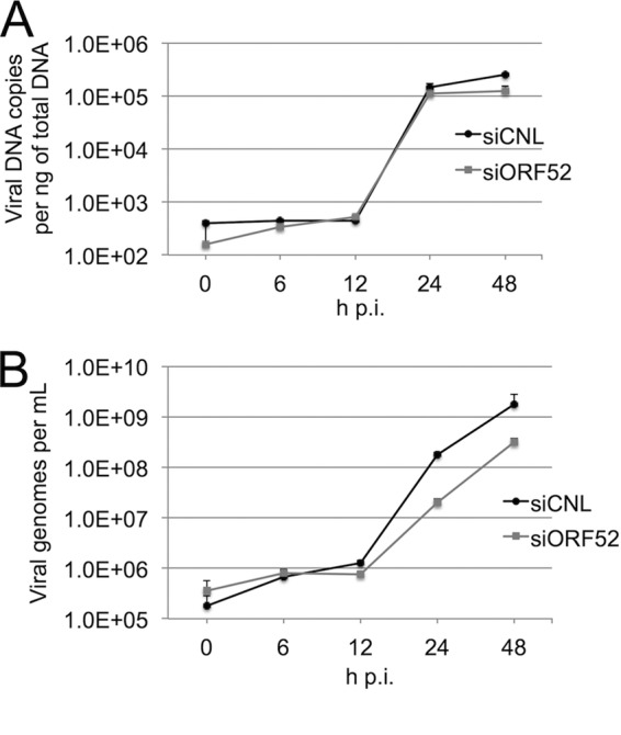
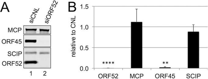
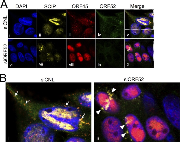
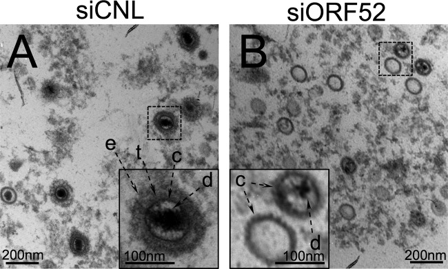
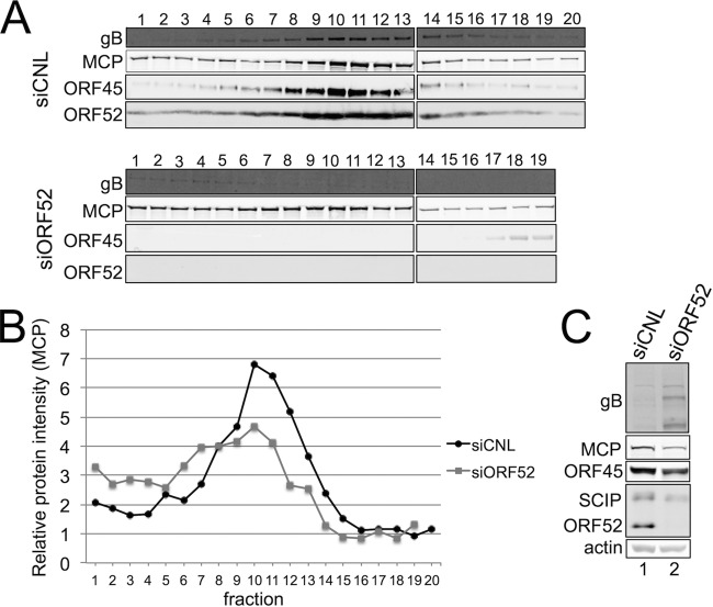
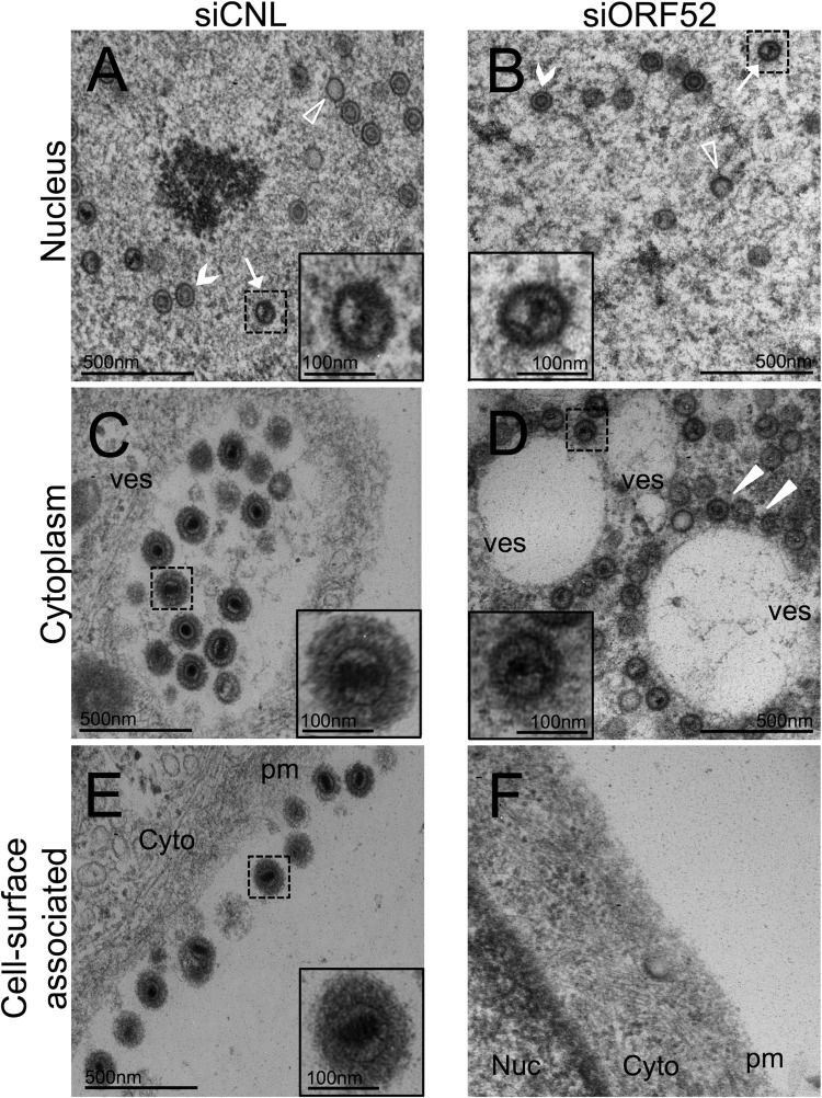
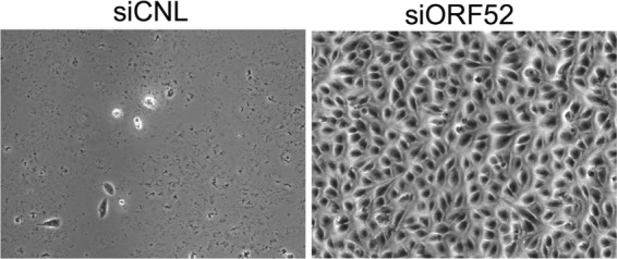
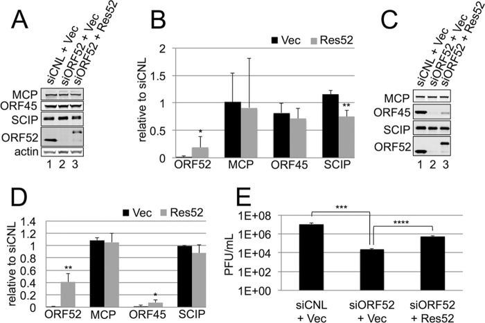
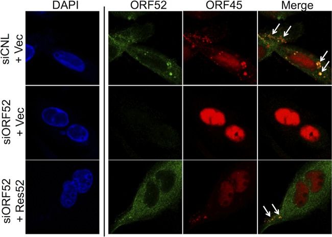
The tegument layer of herpesviruses comprises a collection of proteins that is unique to each viral species. In rhesus monkey rhadinovirus (RRV), a close relative of the human oncogenic pathogen Kaposi's sarcoma-associated herpesvirus, ORF52 is a highly abundant tegument protein tightly associated with the capsid. We now report that ORF52 knockdown during RRV infection of rhesus fibroblasts led to a greater than 300-fold reduction in the viral titer by 48 h but had little effect on the number of released particles and caused only modest reductions in the levels of intracellular viral genomic DNA and no appreciable change in viral DNA packaging into capsids. These data suggested that the lack of ORF52 resulted in the production and release of defective particles. In support of this interpretation, transmission electron microscopy (TEM) revealed that without ORF52, capsid-like particles accumulated in the cytoplasm and were unable to enter egress vesicles, where final tegumentation and envelopment normally occur. TEM also demonstrated defective particles in the medium that closely resembled the accumulating intracellular particles, having neither a full tegument nor an envelope. The disruption in tegument formation from ORF52 suppression, therefore, prevented the incorporation of ORF45, restricting its subcellular localization to the nucleus and appearing, by confocal microscopy, to inhibit particle transport toward the periphery. Ectopic expression of small interfering RNA (siRNA)-resistant ORF52 was able to partially rescue all of these phenotypic changes. In sum, our results indicate that efficient egress of maturing virions and, in agreement with studies on murine gammaherpesvirus 68 (MHV-68), complete tegumentation and secondary envelopment are dependent on intact ORF52.

Importance: The tegument, or middle layer, of herpesviruses comprises both viral and cellular proteins that play key roles in the viral life cycle. A subset of these proteins is present only within members of one of the three subfamilies (alphaherpesviruses, betaherpesviruses, or gammaherpesviruses) of Herpesviridae. In this report, we show that the gammaherpesvirus-specific tegument protein ORF52 is critical for maturation of RRV, the closest relative of Kaposi's sarcoma-associated herpesvirus (KSHV) (a human cancer-causing pathogen) that has undergone this type of analysis. Without ORF52, the nascent subviral particles are essentially stuck in maturation limbo, unable to acquire the tegument or outer (envelope) layers. This greatly attenuates infectivity. Our data, together with earlier work on a murine homolog, as well as a more distantly related human homolog, provide a more complete understanding of how early protein interactions involving virus-encoded tegument proteins are critical for virus assembly and are also, therefore, potentially attractive therapeutic targets.


Figures

FIG 1
FIG 1
Efficient knockdown of ORF52 significantly decreased the RRV titer with only minimal effects on intracellular levels of other structural viral proteins. (A) Immunoblot analysis of cell lysates from RhFs transfected with siCNL (lane 1) or siORF52 (lane 2) and then infected with RRV 24 h later at an MOI of 5. Cells were harvested 48 h p.i., and immunoblotting was performed, probing for the viral tegument proteins ORF52 and ORF45, the capsid proteins MCP and SCIP, and actin to normalize for loading differences. (B) Intracellular levels of the indicated viral structural proteins after siORF52 treatment relative to siCNL in RRV-infected RhFs. The data represent the means and SD of 6 individual experiments. (C) Viral titers in the medium 48 h p.i. from RhFs treated with siCNL or siORF52 were determined in 6 different experiments by viral plaque assay. The values are means and SD. ****, P < 0.0001.
FIG 2
FIG 2
Intracellular viral DNA production and packaging within particles appeared to be independent of ORF52. (A) RhFs were transfected with siCNL or siORF52 and infected 24 h later with RRV at an MOI of 5. At the time points indicated, cells were collected and total intracellular DNA was isolated and purified. Viral DNA was quantified by SYBR green qPCR with primers to the RRV ORF45 coding region. (B) RhFs were transfected with siCNL or siORF52 and infected 24 h later with RRV at an MOI of 5. At the time points indicated, particles were treated with DNase, followed by PK, and viral DNA was isolated, purified, and quantified by SYBR green qPCR with primers for RRV ORF45. The values are the means and SD of 2 or 3 qPCR replicates from 3 separate experiments, with only the positive component of the errors shown for increased graphical clarity.
FIG 3
FIG 3
In the absence of ORF52, RRV infection led to release of immature particles lacking tegument. (A) RhFs were transfected with siCNL (lane 1) or siORF52 (lane 2) and then infected 24 h later with RRV at an MOI of 5. Forty-eight hours p.i., the medium was collected and concentrated over a 20% sucrose cushion to isolate particles, and equal volumes of medium were separated by SDS-PAGE and immunoblotted for MCP, ORF45, SCIP, and ORF52. (B) Effect of siORF52 relative to siCNL on the levels of the indicated viral proteins within the released viral particles. The data represent the means and SD of 6 individual experiments. **, P < 0.01; ****, P < 0.0001.
FIG 4
FIG 4
ORF52 knockdown restricted ORF45 to the nucleus. (A) RhFs were reverse transfected with siCNL (i to v) or siORF52 (vi to x) and plated on coverslips. Twenty-four hours later, the cells were infected with RRV at an MOI of 5, and then at 48 h p.i., the cells were fixed and stained with conjugated anti-SCIP, anti-ORF52, and anti-ORF45 antibodies, as indicated, followed by the secondary antibody Alexa Fluor 647 goat anti-mouse or Alexa Fluor 555 goat anti-rabbit. The cells were stained with DAPI. (B) Boxed areas in panel A, v and x, magnified (3×). The arrows in siCNL (i) point to punctate orange staining (due to ORF45-ORF52 colocalization) in the cytoplasm, consistent with tegumented viral particles during maturation/egress. The arrowheads in siORF52 (ii) point to yellow cytoplasmic staining consistent with SCIP antibody reactivity in the absence of ORF45 and ORF52 reactivity and suggesting untegumented cytoplasmic capsids.
FIG 5
FIG 5
TEM images of the concentrated medium from RRV-infected RhFs after ORF52 knockdown demonstrated release of untegumented and unenveloped capsid-like particles, many of which lacked DNA. RhFs were transfected with siCNL or siORF52 and infected 24 h later with RRV at an MOI of 5. Forty-eight hours p.i., the medium was removed and layered over a sucrose cushion prior to centrifugation. Pelleted particles from the medium were fixed and examined by TEM at ×30,000 magnification. In contrast to the siCNL sample (A), which contained mature virions with envelope (e) and tegument (t) surrounding a capsid (c), particles from siORF52-treated RhFs (B) appeared to lack both envelope and tegument. d, encapsidated viral DNA. The inset images are expanded (3×) from the original images (dashed boxes).
FIG 6
FIG 6
Knockdown of ORF52 leads to release of capsids lacking envelope and tegument. RhFs were transfected with either siCNL or siORF52. After 24 h, the cells were infected with RRV at an MOI of 5 and then harvested 48 h p.i. (A) Medium was collected and concentrated over a 20% sucrose cushion to isolate particles and subsequently layered over a 20 to 60% sucrose gradient. Fractions were collected by bottom puncture, and equal volumes of medium were separated by SDS-PAGE and immunoblotted for gB, MCP, ORF45, and ORF52. (B) Levels of the viral capsid protein MCP present in each fraction. (C) Immunoblot analysis of cell lysates from infected RhFs transfected with siCNL (lane 1) or siORF52 (lane 2). The blots were probed for the viral tegument proteins ORF52 and ORF45, the capsid proteins MCP and SCIP, the glycoprotein gB, and actin to normalize for loading differences.
FIG 7
FIG 7
TEM of RRV-infected cells following ORF52 knockdown showed a block in tegumentation and secondary envelopment, leading to perivesicular accumulation of capsids, as well as a lack of cell surface-associated virions. RhFs were transfected with siCNL or siORF52 and infected as described in the legend to Fig. 5. Forty-eight hours p.i., the cells were fixed and examined by TEM at ×40,000 magnification. (A and B) The nuclei of siCNL-treated (A) and siORF52-treated (B) cells contained A (empty) capsids (arrowheads), B capsids containing scaffold (chevrons), and C capsids with DNA (arrows). (C) The cytoplasm of siCNL cells contained large vesicles (ves) filled with multiple tegumented and enveloped mature virions. (D) In contrast, the cytoplasm of siORF52-treated cells contained untegumented, unenveloped capsids that were juxtaposed to, but not within, vesicles (the arrowheads indicate capsids surrounding a vesicle). (E and F) Multiple extracellular plasma membrane (pm)-associated virions were present in siCNL cells (E), but not in siORF52 samples (F). The inset images are expanded (3×) from the original images. Cyto, cytoplasm; nuc, nucleus.
FIG 8
FIG 8
Knockdown of ORF52 prevents cell-to-cell particle spread. RhFs were transfected with either siCNL or siORF52. Twenty-four hours later, the cells were infected with RRV at an MOI of 0.1, and overlay medium containing methylcellulose was added to inhibit dissemination of released virions while allowing cell-to-cell spread. The plates were examined by phase microscopy 7 days postinfection at ×100 magnification.
FIG 9
FIG 9
Complementation of ORF52 knockdown with exogenous siRNA-resistant ORF52 partially rescued the wild-type phenotype. RhFs transfected with either empty myc-tagged vector (Vec) or siORF52-resistant myc-tagged ORF52 (Res52) were reverse transfected 24 h later with siCNL or siORF52. After an additional 24 h, the cells were infected with RRV at an MOI of 5 and then harvested 48 h p.i. (A) Immunoblot analysis of cell lysates from infected RhFs first transfected with siCNL plus Vec (lane 1), siORF52 plus Vec (lane 2), or siORF52 plus Res52 (lane 3). The protein blots were incubated with antibodies specific for the viral tegument proteins ORF45 and ORF52, the capsid proteins MCP and SCIP, and cellular actin to normalize for loading differences. (B) Levels of the indicated viral proteins after siORF52 treatment with Vec or Res52 relative to siCNL in infected RhFs. The data represent the means and SD of 4 individual experiments. (C) Immunoblot analysis of concentrated medium collected from RhFs transfected with siCNL plus Vec (lane 1), siORF52 plus Vec (lane 2), or siORF52 plus Res52 (lane 3). Protein blots from the medium were probed for ORF45, ORF52, MCP, and SCIP. (D) Levels of the indicated viral proteins in released particles after siORF52 treatment following transfection with Vec or Res52 relative to those in particles released from siCNL-treated cells. The data represent the means and SD of 4 individual experiments. (B and D) P values were calculated by comparing knockdown (black bars) to rescue (gray bars). (E) Viral titers in the medium from each condition were determined by viral plaque assays of 4 individual experiments. The values are means and SD. *, P < 0.05; **, P < 0.01; ***, P < 0.001; ****, P < 0.0001.
FIG 10
FIG 10
Exogenous ORF52 rescues formation and release of tegumented and enveloped virions. RhFs transfected with either empty myc-tagged vector (Vec) or siORF52-resistant myc-tagged ORF52 (Res52) were transfected 24 h later with siCNL or siORF52 and then, after an additional 24 h, were infected at an MOI of 5. Forty-eight hours later, the cells were fixed and examined by TEM at ×40,000 magnification. The nuclei of cells treated with (A) siCNL plus vec, (B) siORF52 plus vec, (C) or siORF52 plus Res52 all contained A (empty) (arrowheads), B scaffold-containing (chevrons), and C (arrows) capsids. (D and E) The cytoplasm of siCNL plus Vec cells (D) contained large vesicles (ves) filled with multiple tegumented and enveloped virions; the cytoplasm of siORF52 plus Vec cells (E) contained untegumented particles, many of which surrounded but were not within vesicles. (F) siORF52 plus Res52 cells showed virions within cytoplasmic vesicles. (G to I) In cells treated with siORF52 plus Vec (H), cell-associated virions are absent, in contrast to cells treated with siCNL plus vec (G) or siORF52 plus Res52 (I). (J to L) Cells treated with siORF52 plus Vec release particles that appear to be capsids, lacking tegument and envelope (K), in contrast to cells treated with siCNL plus Vec (J) and siORF52 plus Res52 (L), which release intact, mature virions. The inset images are expanded (2×) from the original images.
FIG 11
FIG 11
Complementation with siRNA-resistant ORF52 partially restored cytoplasmic subcellular localization of ORF45. RhFs transfected with either empty myc-tagged vector (Vec) or siORF52-resistant myc-tagged ORF52 (Res52) were reverse transfected 24 h later on coverslips with siCNL (top row) or siORF52 (middle and bottom rows) and then, after 24 h, infected with RRV at an MOI of 5 and fixed 48 h later. The cells were stained with anti-ORF52 and anti-ORF45 antibodies, followed by the secondary antibodies Alexa Fluor 488 goat anti-mouse and Alexa Fluor 555 goat anti-rabbit, respectively. The cells were then stained with DAPI. The arrows highlight overlapping signals and colocalization of ORF52 and ORF45 in the cytoplasm.

Similar articles

Cited by

References

    1. Cesarman E, Chang Y, Moore PS, Said JW, Knowles DM. 1995. Kaposi's sarcoma-associated herpesvirus-like DNA sequences in AIDS-related body-cavity-based lymphomas. N. Engl. J. Med. 332:1186–1191. 10.1056/NEJM199505043321802 - DOI - PubMed
    1. Chang Y, Cesarman E, Pessin MS, Lee F, Culpepper J, Knowles DM, Moore PS. 1994. Identification of herpesvirus-like DNA sequences in AIDS-associated Kaposi's sarcoma. Science 266:1865–1869. 10.1126/science.7997879 - DOI - PubMed
    1. Cesarman E, Moore PS, Rao PH, Inghirami G, Knowles DM, Chang Y. 1995. In vitro establishment and characterization of two acquired immunodeficiency syndrome-related lymphoma cell lines (BC-1 and BC-2) containing Kaposi's sarcoma-associated herpesvirus-like (KSHV) DNA sequences. Blood 86:2708–2714 - PubMed
    1. Boshoff C, Chang Y. 2001. Kaposi's sarcoma-associated herpesvirus: a new DNA tumor virus. Annu. Rev. Med. 52:453–470. 10.1146/annurev.med.52.1.453 - DOI - PubMed
    1. Mettenleiter TC, Klupp BG, Granzow H. 2009. Herpesvirus assembly: an update. Virus Res. 143:222–234. 10.1016/j.virusres.2009.03.018 - DOI - PubMed

Publication types

LinkOut - more resources