. 2014 Aug;88(16):9141-52.
doi: 10.1128/JVI.00619-14. Epub 2014 Jun 4.

HIV-1 Vpr redirects host ubiquitination pathway

Affiliations

HIV-1 Vpr redirects host ubiquitination pathway

Sakshi Arora et al. J Virol. 2014 Aug.

Abstract

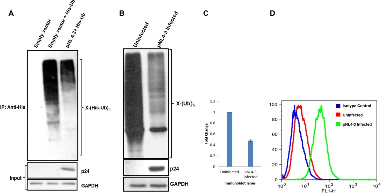
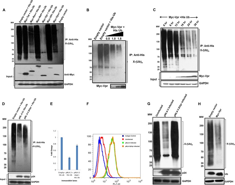
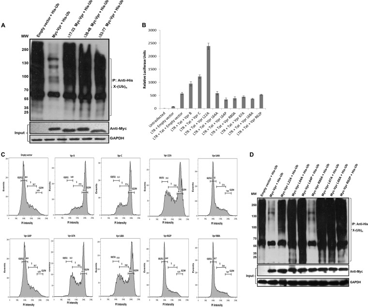
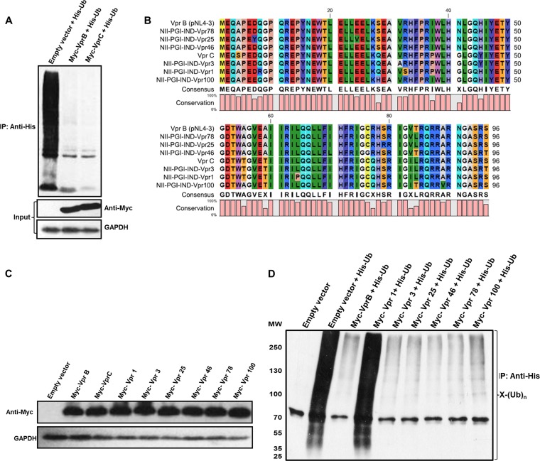
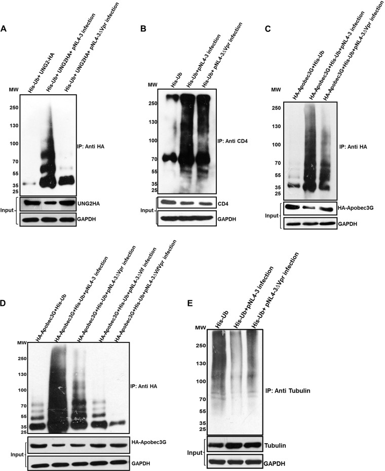
HIV-1 modulates key host cellular pathways for successful replication and pathogenesis through viral proteins. By evaluating the hijacking of the host ubiquitination pathway by HIV-1 at the whole-cell level, we now show major perturbations in the ubiquitinated pool of the host proteins post-HIV-1 infection. Our overexpression- and infection-based studies of T cells with wild-type and mutant HIV-1 proviral constructs showed that Vpr is necessary and sufficient for reducing whole-cell ubiquitination. Mutagenic analysis revealed that the three leucine-rich helical regions of Vpr are critical for this novel function of Vpr, which was independent of its other known cellular functions. We also validated that this effect of Vpr was conserved among different subtypes (subtypes B and C) and circulating recombinants from Northern India. Finally, we establish that this phenomenon is involved in HIV-1-mediated diversion of host ubiquitination machinery specifically toward the degradation of various restriction factors during viral pathogenesis.

Importance: HIV-1 is known to rely heavily on modulation of the host ubiquitin pathway, particularly for counteraction of antiretroviral restriction factors, i.e., APOBEC3G, UNG2, and BST-2, etc.; viral assembly; and release. Reports to date have focused on the molecular hijacking of the ubiquitin machinery by HIV-1 at the level of E3 ligases. Interaction of a viral protein with an E3 ligase alters its specificity to bring about selective protein ubiquitination. However, in the case of infection, multiple viral proteins can interact with this multienzyme pathway at various levels, making it much more complicated. Here, we have addressed the manipulation of ubiquitination at the whole-cell level post-HIV-1 infection. Our results show that HIV-1 Vpr is necessary and sufficient to bring about the redirection of the host ubiquitin pathway toward HIV-1-specific outcomes. We also show that the three leucine-rich helical regions of Vpr are critical for this effect and that this ability of Vpr is conserved across circulating recombinants. Our work, the first of its kind, provides novel insight into the regulation of the ubiquitin system at the whole-cell level by HIV-1.


Figures

FIG 1
FIG 1
pNL4-3 transfection or infection reduces whole-cell ubiquitination. (A) HEK 293T cells were cotransfected with plasmids encoding His-Ub and pCMV-Myc or pNL4-3. After 36 h, cells were treated with MG132 for 8 h, followed by Ni-NTA pulldown. Immunoblot analysis was done, and whole-cell ubiquitination was probed by using an anti-His antibody. p24 was indicative of infection. GAPDH was used as a loading control (input). IP, immunoprecipitation. (B) Jurkat cells were infected with pNL4-3 viral supernatants for 4 h, and after 36 h, they were treated with MG132 for 8 h. The lysate was prepared in RIPA buffer containing 1× protease inhibitor and 5 mM N-ethylmaleimide. Immunoblot analysis was done, and whole-cell ubiquitination was probed by using an anti-Ub antibody. p24 was indicative of infection. GAPDH was used as a loading control. (C) Quantitation of ubiquitination for the immunoblot shown in panel B. (D) Jurkat cells were evaluated for the extent of HIV-1 infection by intracellular p24 staining with primary p24 mouse antibody (NIH) followed by secondary anti-mouse antibody (FITC conjugated). FITC staining in flow cytometer channel 1 (FL1) was analyzed; H indicates height.
FIG 2
FIG 2
Vpr is sufficient to reduce whole-cell ubiquitination. (A) HEK 293T cells were cotransfected with His-Ub and pCMV-Myc or each viral accessory/regulatory gene (Myc-TatB/RevB/NefB/VprB/VifB/VpuB). After 36 h, cells were treated with MG132 for 8 h, followed by Ni-NTA pulldown. Immunoblot analysis was done, and whole-cell ubiquitination was probed by using an anti-His antibody. MW, molecular weight marker (in thousands). (B) Increasing doses of Myc-VprB were cotransfected with His-Ub, and immunoblotting was done as described above for panel A. The level of VprB is shown as the input. (C) A time course assay was done. After 12 h of transfection, Vpr was detected in lysates, and whole-cell ubiquitination was probed at the same time intervals. (D) HEK 293T cells were cotransfected with His-Ub and pNL4-3/pNL4-3Δvpr, and whole-cell ubiquitination was probed as described above for panel A. (E) Quantitation of ubiquitination for the immunoblot shown in panel D. (F and G) Jurkat cells were infected with pNL4-3/pNL4-3Δvpr viral supernatants for 4 h, and after 36 h, they were treated with MG132 for 8 h. Cells were divided; one set was used for intracellular p24 staining (as described in the legend of Fig. 1D), the other set was lysed, and whole-cell ubiquitination was probed by using an anti-Ub antibody. GAPDH was used as a loading control. (H) Endogenous ubiquitination was probed by using an anti-Ub antibody after overexpression of Myc-VprB in HEK 293T cells. Levels of free ubiquitin are also shown. GAPDH was used as a loading control.
FIG 3
FIG 3
The three helical regions of Vpr critical for reducing whole-cell ubiquitination. (A) The three helices were independently deleted (Δ17-33, Δ38-48, and Δ53-77 Myc-tagged Vpr deletions). Expression was checked in HEK 293T cells, and each deletion construct was then cotransfected with His-Ub. After 36 h, MG132 treatment was given for 8 h, and ubiquitinated proteins were enriched by using Ni-NTA beads (as described in Materials and Methods). Immunoblotting was done by using an anti-His antibody to probe whole-cell ubiquitination. (B) wt Vpr and point mutants were checked for their abilities to transactivate the LTR by using the LTR-luc construct (as described in Materials and Methods). The results are representative of three independent experiments. (C) wt Vpr and point mutants were checked for their abilities to cause G2/M arrest. HEK 293T cells were collected at 48 h posttransfection and were stained with propidium iodide as described in Materials and Methods. (D) wt Vpr and point mutants were then cotransfected with His-Ub in HEK 293T cells. After 36 h, MG132 treatment was given for 8 h, and ubiquitinated proteins were enriched by using Ni-NTA beads (as described in Materials and Methods). Immunoblotting was done by using an anti-His antibody to probe whole-cell ubiquitination. GAPDH was used as a loading control.
FIG 4
FIG 4
Comparison of HIV-1 subtypes B and C and natural variants. (A) HEK 293T cells were cotransfected with Myc-VprB/VprC and His-Ub. After 36 h, MG132 treatment was given for 8 h, and ubiquitinated proteins were enriched by using Ni-NTA beads (as described in Materials and Methods). Immunoblotting was done by using an anti-His antibody to probe whole-cell ubiquitination. Levels of Myc-VprB and subtype C are shown as the input. (B) Protein sequence alignment of HIV-1 VprB, VprC, and 6 samples. (C) The expression levels of 6 variant samples (Vpr-1, -3, -25, -46, -78, and -100), Myc-VprB, and Myc-VprC were checked in HEK 293T cells and normalized. (D) HEK 293T cells were cotransfected with Myc-VprB, 6 variant samples (Vpr-1, -3, -25, -46, -78, and -100), and His-Ub. After 36 h, MG132 treatment was given for 8 h, and ubiquitinated proteins were enriched by using Ni-NTA beads (as described in Materials and Methods). Immunoblotting was done by using an anti-His antibody to probe whole-cell ubiquitination. GAPDH was used as a loading control.
FIG 5
FIG 5
Ubiquitination and degradation of different proteins. (A) HEK 293T cells were cotransfected with HA-UNG2 and His-Ub. After 12 h, the cells were infected with equal multiplicities of infection of pNL4-3/pNL4-3Δvpr VSV-G-pseudotyped virus. After 36 h, MG132 treatment was given for 8 h, and ubiquitinated proteins were enriched by using Ni-NTA beads. Immunoblot analysis was done by using an anti-HA antibody. (B) TZM-bl cells were transfected with His-Ub. After 12 h, the cells were infected with equal multiplicities of infection of pNL4-3/pNL4-3Δvpr VSV-G-pseudotyped virus. After 36 h, MG132 treatment was given for 8 h, and ubiquitinated proteins were enriched by using Ni-NTA beads. Immunoblot analysis was done by using anti-CD4 antibody. (C) HEK 293T cells were cotransfected with HA-APOBEC3G and His-Ub. After 12 h, the cells were infected with equal multiplicities of infection of pNL4-3/pNL4-3Δvpr VSV-G-pseudotyped virus. After 36 h, MG132 treatment was given for 8 h, and ubiquitinated proteins were enriched by using Ni-NTA beads. Immunoblot analysis was done by using an anti-HA antibody. (D) HEK 293T cells were cotransfected with HA-APOBEC3G and His-Ub. After 12 h, the cells were infected with equal multiplicities of infection of pNL4-3/pNL4-3Δvpr/pNL4-3Δvif/pNL4-3ΔvifΔvpr VSV-G-pseudotyped virus. After 36 h, MG132 treatment was given for 8 h, and ubiquitinated proteins were enriched by using Ni-NTA beads. Immunoblot analysis was done by using an anti-HA antibody. (E) HEK 293T cells were transfected with His-Ub. After 12 h, the cells were infected with equal multiplicities of infection of pNL4-3/pNL4-3Δvpr VSV-G-pseudotyped virus. After 36 h, MG132 treatment was given for 8 h, and ubiquitinated proteins were enriched by using Ni-NTA beads. Immunoblot analysis was done by using an anti-tubulin antibody. Levels of proteins are shown as the input (without MG132 treatment). GAPDH was used as a loading control.

Similar articles

Cited by

References

    1. Clapham PR, McKnight A. 2001. HIV-1 receptors and cell tropism. Br. Med. Bull. 58:43–59. 10.1093/bmb/58.1.43 - DOI - PubMed
    1. Cummins NW, Badley AD. 2010. Mechanisms of HIV-associated lymphocyte apoptosis: 2010. Cell Death Dis. 1:e99. 10.1038/cddis.2010.77 - DOI - PMC - PubMed
    1. Cullen BR, Greene WC. 1990. Functions of the auxiliary gene products of the human immunodeficiency virus type 1. Virology 178:1–5. 10.1016/0042-6822(90)90373-Y - DOI - PubMed
    1. Malim MH, Emerman M. 2008. HIV-1 accessory proteins—ensuring viral survival in a hostile environment. Cell Host Microbe 3:388–398. 10.1016/j.chom.2008.04.008 - DOI - PubMed
    1. Kirchhoff F. 2010. Immune evasion and counteraction of restriction factors by HIV-1 and other primate lentiviruses. Cell Host Microbe 8:55–67. 10.1016/j.chom.2010.06.004 - DOI - PubMed

Publication types

MeSH terms