

Simultaneous gene deletion of gata4 and gata6 leads to early disruption of follicular development and germ cell loss in the murine ovary
- PMID: 24899573
- PMCID: PMC4434962
- DOI: 10.1095/biolreprod.113.117002
Simultaneous gene deletion of gata4 and gata6 leads to early disruption of follicular development and germ cell loss in the murine ovary
Abstract
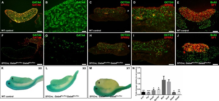
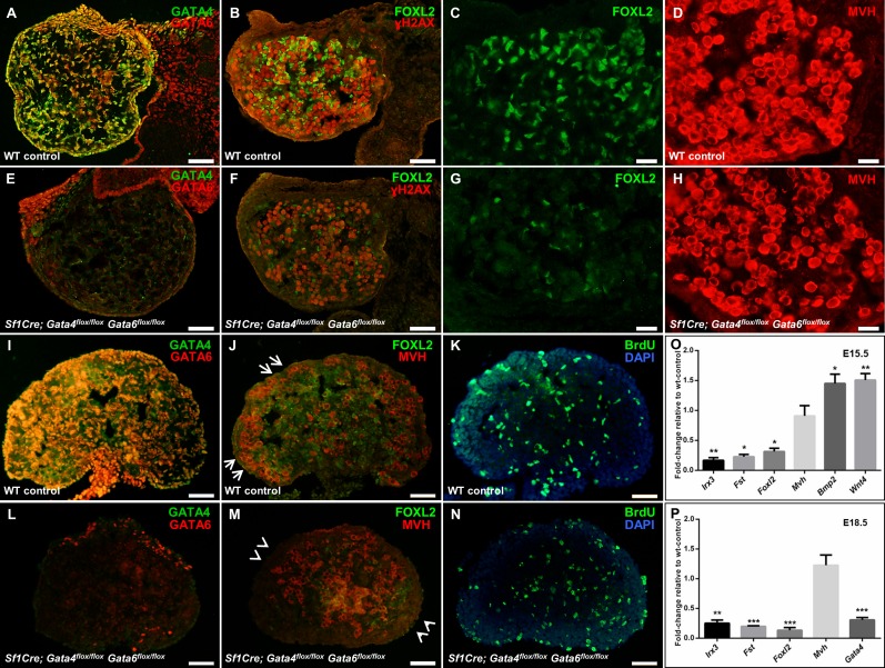
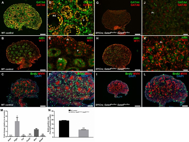
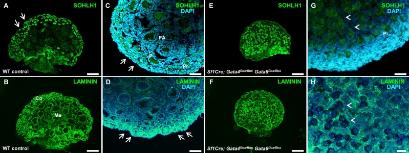
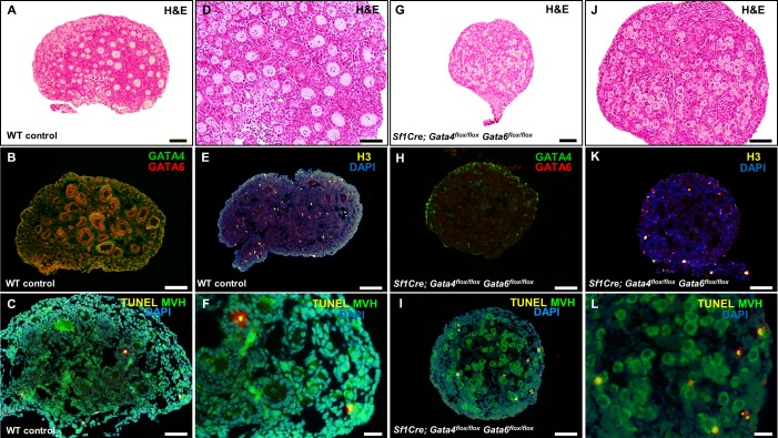
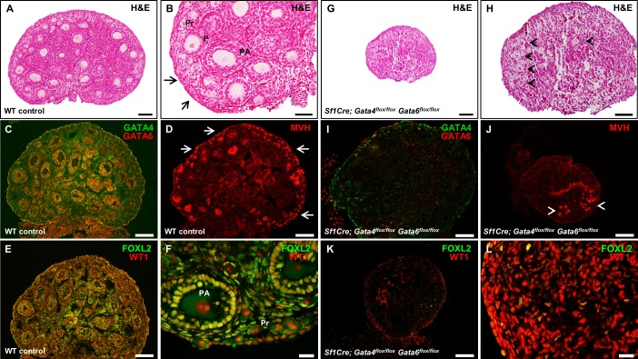
Granulosa cell formation and subsequent follicular assembly are important for ovarian development and function. Two members of the GATA family of transcription factors, GATA4 and GATA6, are expressed in ovarian somatic cells early in development, and their importance in adult ovarian function has been recently highlighted. In this study, we demonstrated that the embryonic loss of Gata4 and Gata6 expression within the ovary results in a strong down-regulation of genes involved in the ovarian developmental pathway (Fst and Irx3) as well as diminished expression of the pregranulosa and granulosa cell markers SPRR2 and FOXL2, respectively. Postnatal ovaries deficient in both Gata genes show impaired somatic cell proliferation and arrested follicular development at the primordial stage, where oocytes are either enclosed by one layer of squamous granulosa cells or remain in germ cell nests/clusters. Furthermore, germ cell nests and primordial follicles are predominantly localized to the central region of the Sf1Cre; Gata4(flox/flox) Gata6(flox/flox) ovaries, where the boundary between the medulla and cortex is almost nonexistent. Lastly, most of the oocytes are lost early in development in conditional double mutant ovaries, which confirms the importance of normally differentiated granulosa cells as supporting cells for oocyte survival. Thus, both GATA4 and GATA6 proteins are fundamental regulators of granulosa cell differentiation and proliferation, and consequently of proper follicular assembly during normal ovarian development and function.
Keywords: differentiation; granulosa cells; ovarian development.
© 2014 by the Society for the Study of Reproduction, Inc.
Figures
Comment in
-
The GATA-keepers of ovarian development and folliculogenesis.Biol Reprod. 2014 Aug;91(2):38. doi: 10.1095/biolreprod.114.122499. Epub 2014 Jul 2. Biol Reprod. 2014. PMID: 24990805 No abstract available.
Similar articles
-
Adrenal Development in Mice Requires GATA4 and GATA6 Transcription Factors.Endocrinology. 2015 Jul;156(7):2503-17. doi: 10.1210/en.2014-1815. Epub 2015 May 1. Endocrinology. 2015. PMID: 25933105 Free PMC article.
-
Combined loss of the GATA4 and GATA6 transcription factors in male mice disrupts testicular development and confers adrenal-like function in the testes.Endocrinology. 2015 May;156(5):1873-86. doi: 10.1210/en.2014-1907. Epub 2015 Feb 10. Endocrinology. 2015. PMID: 25668066 Free PMC article.
-
GATA4 and GATA6 silencing in ovarian granulosa cells affects levels of mRNAs involved in steroidogenesis, extracellular structure organization, IGF-I activity, and apoptosis.Endocrinology. 2013 Dec;154(12):4845-58. doi: 10.1210/en.2013-1410. Epub 2013 Sep 24. Endocrinology. 2013. PMID: 24064357 Free PMC article.
-
GATA Regulation and Function During the Ovarian Life Cycle.Vitam Horm. 2018;107:193-225. doi: 10.1016/bs.vh.2018.01.014. Epub 2018 Feb 13. Vitam Horm. 2018. PMID: 29544631 Free PMC article. Review.
-
[The GATA family in reproduction].Zhonghua Nan Ke Xue. 2009 Oct;15(10):932-6. Zhonghua Nan Ke Xue. 2009. PMID: 20112745 Review. Chinese.
Cited by
-
A Hormone That Lost Its Receptor: Anti-Müllerian Hormone (AMH) in Zebrafish Gonad Development and Sex Determination.Genetics. 2019 Oct;213(2):529-553. doi: 10.1534/genetics.119.302365. Epub 2019 Aug 9. Genetics. 2019. PMID: 31399485 Free PMC article.
-
Adrenal Development in Mice Requires GATA4 and GATA6 Transcription Factors.Endocrinology. 2015 Jul;156(7):2503-17. doi: 10.1210/en.2014-1815. Epub 2015 May 1. Endocrinology. 2015. PMID: 25933105 Free PMC article.
-
Transcript abundance of stromal and thecal cell related genes during bovine ovarian development.PLoS One. 2019 Mar 11;14(3):e0213575. doi: 10.1371/journal.pone.0213575. eCollection 2019. PLoS One. 2019. PMID: 30856218 Free PMC article.
-
SP1 governs primordial folliculogenesis by regulating pregranulosa cell development in mice.J Mol Cell Biol. 2020 Apr 24;12(3):230-244. doi: 10.1093/jmcb/mjz059. J Mol Cell Biol. 2020. PMID: 31282930 Free PMC article.
-
Transposable elements acquire time- and sex-specific transcriptional and epigenetic signatures along mouse fetal gonad development.Front Cell Dev Biol. 2024 Jan 12;11:1327410. doi: 10.3389/fcell.2023.1327410. eCollection 2023. Front Cell Dev Biol. 2024. PMID: 38283992 Free PMC article.
References
-
- Molkentin JD. The zinc finger-containing transcription factors GATA-4, -5, and -6. Ubiquitously expressed regulators of tissue-specific gene expression. J Biol Chem. 2000;275:38949–38952. - PubMed
-
- Zaytouni T, Efimenko EE, Tevosian SG. GATA transcription factors in the developing reproductive system. Adv Genet. 2011;76:93–134. - PubMed
-
- Siggers P, Smith L, Greenfield A. Sexually dimorphic expression of Gata-2 during mouse gonad development. Mech Dev. 2002;111:159–162. - PubMed
Publication types
MeSH terms
Substances
Grants and funding
LinkOut - more resources
Full Text Sources
Other Literature Sources
Molecular Biology Databases
Miscellaneous
