. 2014 Aug 7;124(6):924-35.
doi: 10.1182/blood-2014-01-549162. Epub 2014 Jun 4.

A motif in LILRB2 critical for Angptl2 binding and activation

Affiliations

A motif in LILRB2 critical for Angptl2 binding and activation

Mi Deng et al. Blood. .

Abstract

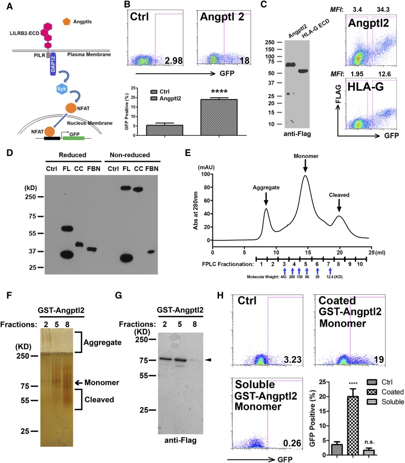
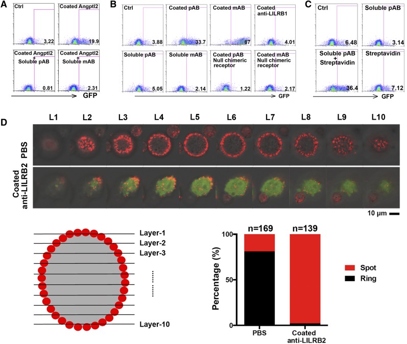
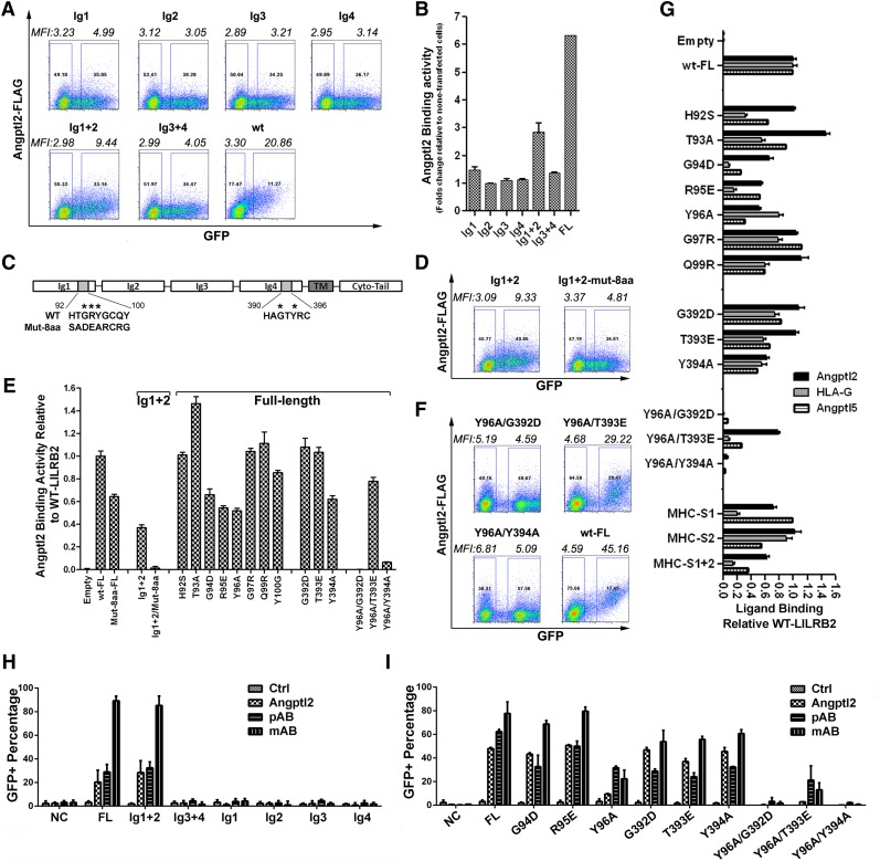
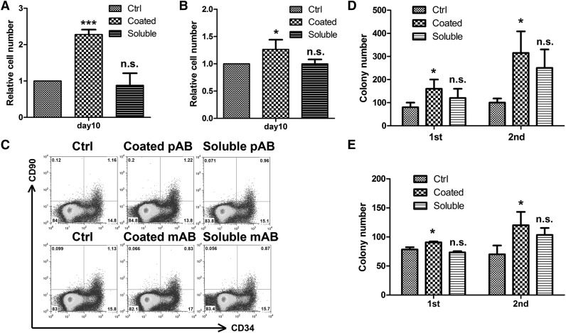
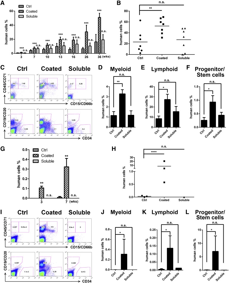
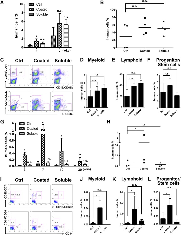
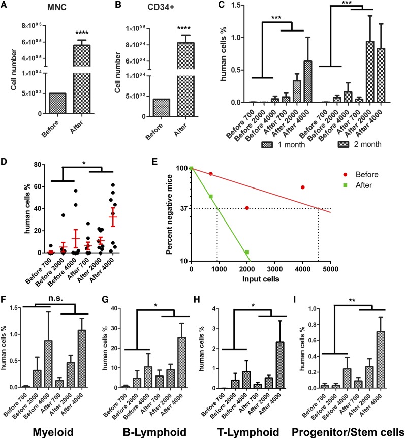
A better understanding of the interaction between extrinsic factors and surface receptors on stem cells will greatly benefit stem cell research and applications. Recently, we showed that several angiopoietin-like proteins (Angptls) bind and activate the immune inhibitory receptor human leukocyte immunoglobulin (Ig)-like receptor B2 (LILRB2) to support ex vivo expansion of hematopoietic stem cells (HSCs) and leukemia development. However, the molecular basis for the interaction between Angptls and LILRB2 was unclear. Here, we demonstrate that Angptl2 expressed in mammalian cells forms high-molecular-weight species and that ligand multimerization is required for activation of LILRB2 for downstream signaling. A novel motif in the first and fourth Ig domains of LILRB2 was identified that is necessary for the receptor to be bound and activated by Angptl2. The binding of Angptl2 to LILRB2 is more potent than and not completely overlapped with the binding of another ligand, HLA-G. Immobilized anti-LILRB2 antibodies induce a more potent activation of LILRB2 than Angptl2, and we developed a serum-free culture containing defined cytokines and immobilized anti-LILRB2 that supports a net expansion of repopulating human cord blood HSCs. Our elucidation of the mode of Angptl binding to LILRB2 enabled the development of a new approach for ex vivo expansion of human HSCs.


Figures

Figure 1
Figure 1
HMW Angptl2 activates LILRB2 signaling. (A) Schematic of the chimeric LILRB2 receptor reporter system. (B) Representative flow cytometric profiles and summary showing that the Angptl2-conditioned medium stimulates GFP induction in the LILRB2 chimeric reporter system. The condition media of empty-vector–transfected HEK-293T cells was used as control. (C) Secreted Angplt2 and HLA-G-ECD in condition medium detected by anti-FLAG antibody in western blotting (left). Representative flow cytometric plots showing that Angptl2 binds to LILRB2 expressed on HEK293T cells better than the same amount of HLA-G-ECD (right). (D) The full-length (FL), CC, and FBN domains obtained from conditioned medium showed distinctive migration in reducing and nonreducing sodium dodecyl sulfate polyacrylamide gel electrophoresis as determined by immunoblotting with anti-M2 Flag antibody. Protein extracted from equivalent amounts of condition media of empty-vector–transfected HEK293T cells was used as control. (E) GST-human Angptl2 purified from bacterial expression system by GST was immediately fractionated through gel-filtration FPLC. The molecular weight was determined by the peaks of apoferritin (443 kDa), amylase (200 kDa), alcohol dehydrogenase (150 kDa), albumin (66 kDa), carbonic anhydrase (29 kDa), and cytochrome c (12.4 kDa), respectively. (F) Equivalent amounts of indicated fractionated samples in FPLC were loaded on 10% native gel. Aggregated, monomeric, and cleaved GST-Angptl2 were visualized by silver staining. (G) Indicated FPLC fractionated samples were examined by western blotting using anti-M2 Flag antibody. The FLAG in cleaved GST-Angptl2 fragments (fraction 8; Figure 1G) could not be detected by western blotting. (H) Chimeric LILRB2 receptor reporter cells were treated with coated or soluble fraction 5 proteins for 48 hours. In coated wells, 5 µg/ml GST-Angptl2 from fraction 5 was precoated onto wells of a 96-well plate for 3 hours at 37°C. An equivalent amount of FPLC buffer was used as control. n.s. indicates not significant; ****P < .0001. Ctrl, control; KD, kilodalton.
Figure 2
Figure 2
Immobilized anti-LILRB2 antibodies activated the chimeric LILRB2 reporter. (A) Representative flow cytometric profiles showing that the GFP induction by immobilized 5 µg/mL Angptl2 was abolished by 5 µg/mL anti-LILRB2 antibody. Chimeric LILRB2 receptor reporter cells were treated with indicated coated Angptl2 with or without soluble anti-LILRB2 pAb or mAb for 48 hours. Phosphate-buffered saline (PBS) was used as control. (B) Representative flow cytometric profiles showing that GFP was induced by immobilized anti-LILRB2 antibodies. Chimeric LILRB2 receptor reporter cells were treated with indicated coated (25 µg/mL in 50 µL PBS) or soluble (5 µg/mL in 250 µL cell culture media) antibodies for 48 hours. The reporter cells not containing chimeric LILRB2 receptor were used as negative control. (C) Representative flow cytometric profiles showing that GFP expression was induced by crosslinked anti-LILRB2 antibodies. Chimeric LILRB2 receptor reporter cells were treated with 10 µg/mL soluble anti-LILRB2 polyclone antibody (pAb) or equivalent crosslinked pAb for 48 hours. Streptavidin alone was used as a negative control. (D) Representative confocal images of LILRB2 chimeric receptor reporter cells with or without coated anti-LILRB2 mAb showing that the distribution of LILRB2 protein on cell plasma membrane. Ten confocal scans from top to bottom of a cell were indicated from layer 1 (L1) to layer 10 (L10). Confocal images of the phase contrast, Cy3 (indicating LILRB2 expression), and GFP (indicating signaling activation) panels were merged. Ctrl, control.
Figure 3
Figure 3
Ig domains 1 and 4 in LILRB2 are critical for Angptl2 binding and signal activation. (A) Representative flow cytometry plots showing Angptl2 binding to full-length, individual Ig domain, Ig1+2, or Ig3+4 of LILRB2 that were expressed on 293T cells; n = 3. (B) Summary of data from panel A. (C) Summary of Angptl2 binding abilities of wild-type (WT) and mutant LILRB2. Indicated mutations are described in supplemental Figure 3B. (C) Schematic of the H*G*Y*C motifs in Ig1 and Ig4 of LILRB2. (D) Summary of Angptl2 binding abilities of WT and mutant Ig1+2 LILRB2. (E) Representative flow cytometry plots showing Angptl2 binding to Ig1+2 and mutant LILRB2. (F) Representative flow cytometry plots showing Angptl2 binding to WT and mutant LILRB2. (G) Comparison of Angptl2, Angptl5, and HLA-G binding abilities of WT and mutant LILRB2. MHC-S indicates HLA-G binding sites; MHC-S1, R59A/Y61A; MHC-S2, W90A/D200A/N202A/Y205A; MHC-S1+2, R59A/Y61A/W90A/D200A/N202A/Y205A. (H) Summary of Angptl2-induced activation of the chimeric receptor reporter system by individual Ig domains, Ig1+2, or Ig3+4 of LILRB2. Indicated reporter cells were treated with 5 µg/mL coated GST-Angptl2 or polyclonal or monoclonal anti-LILRB2 antibodies. At least 3 independent experiments gave the similar results. (I) Summary of Angptl2-induced activation of the chimeric receptor reporter system by WT or mutant LILRB2. Reporter cells were treated with 10 µg/mL coated GST-Angptl2 or polyclonal or monoclonal anti-LILRB2 antibodies. At least 3 independent experiments were performed that gave the similar results. NC, negative control cells that do not express the chimeric receptors.
Figure 4
Figure 4
Immobilized anti-LILRB2 antibodies promote the proliferation of human cord blood cells in vitro. (A) Human CD133+ umbilical cord blood cells were cultured in STF medium with or without the same amounts of coated (25 µg/mL in 50 µL PBS) or soluble (5 µg/mL in 250 µL StemSpan media) anti-LILRB2 pAb. Total cell expansion was assessed after 10 days of culture (n = 3). (B) Human CD133+ umbilical cord blood cells were cultured in STF medium with or without the same amounts of coated (25 µg/mL in 50 µL PBS) or soluble (5 µg/mL in 250 µL StemSpan media) anti-LILRB2 mAb. Total cell expansion was assessed after 10 days of culture (n = 3). (C) Representative flow cytometric profiles showing the frequency of CD34+CD90+ cells after 10 days of culture. (D-E) Expansion of 250 input equivalent human cord blood CD133+ cells treated with or without anti-LILRB2 pAb (D) or mAb (E) were serially plated in CFU medium. Total CFUs were counted after 7 days in culture. *P < .05; ***P < .001. Ctrl, control; n.s., not significant.
Figure 5
Figure 5
Ex vivo expansion of human cord blood CD133+ cells by anti-LILRB2 pAb as determined by NSG transplantation. (A) After 10 days of culture in STF medium with or without same amounts of coated (25 µg/mL in 50 µL PBS) or soluble (5 µg/mL in 250 µL StemSpan media) anti-LILRB2 pAb, expansion of 1 × 10⁴ input equivalent human cord blood CD133+ cells were transplanted into NSG mice (n = 8). Engraftment of human cells (human CD45+) in peripheral blood at indicated weeks are shown***P < .001. (B) Engraftment of human CD45/CD71+ in bone marrow of mice described in panel A at 36 weeks. *P < .05; n = 8. (C) Multilineage contribution of cultured human umbilical cord blood CD133+ cells. Shown are representative flow cytometric profiles of bone marrow cells from 1 primary transplanted mouse of each group. Myeloid, CD45/CD71+CD15/CD66b+; lymphoid, CD19/CD20+; hematopoietic stem/progenitor cells, CD19/CD20CD34+. (D-F) Summary of multilineage contributions from data shown in panel C. *P < .05; **P < .01; n = 8. (G) Engraftment of human CD45+ cells in peripheral blood of secondarily transplanted mice at 3 and 7 weeks posttransplant are shown. **P < .01; n = 3. (H) Engraftments of human cells in bone marrow of secondarily transplanted mice at 8 weeks posttransplant are shown. *P < .05; n = 3. (I) Representative flow cytometric profiles showing multilineage contribution of human umbilical cord blood CD133+ cells in the bone marrow of secondarily transplanted mice at 8 weeks posttransplant. (J-L) Summary of multilineage contributions from data shown in panel I. *P < .05; n = 3. Ctrl, control; n.s., not significant.
Figure 6
Figure 6
Ex vivo expansion of human cord blood CD133+ cells by anti-LILRB2 monoclonal antibody in NSG mice as determined by NSG transplantation. (A) After 10 days culture in STF medium with or without same amounts of coated (25 µg/mL in 250 µL PBS) or soluble (5 µg/mL in 250 µL StemSpan media) anti-LILRB2 mAb, 1 × 10⁴ input equivalent human cord blood CD133+ cells were transplanted into NSG mice. Engraftment of human CD45+ in peripheral blood at 3 and 7 weeks are shown. *P < .05; n = 4. (B) Engraftments of human CD45/CD71+ in bone marrow of mice described in panel A at 8 weeks; n = 4. (C) Multilineage contribution of cultured human umbilical cord blood CD133+ cells. Shown are representative flow cytometric profiles of bone marrow cells from 1 primary transplanted mouse of each group. (D-F) Summary of multilineage contributions based on data shown in panel C; n = 4. (G) Engraftment of human CD45+ cells in peripheral blood of secondarily transplanted mice at 3, 7, 10, and 30 weeks. *P < .05; n = 3. (H) Engraftment of human cells in bone marrow of secondarily transplanted mice at 30 weeks; n = 3. (I) Representative flow cytometric profiles showing multilineage contribution of human umbilical cord blood CD133+ cells in the bone marrow of secondarily transplanted mice at 8 weeks posttransplant. (J-L) Summary of multilineage contributions based on data from panel I. *P < .05; n = 3. Ctrl, control; n.s., not significant.
Figure 7
Figure 7
Net ex vivo expansion of cultured human umbilical cord blood CD133+ cells as determined by limiting dilution analysis. (A-B) Numbers of total nucleated cells (A) and CD34+ cells (B) before and after culture with 25 µg/mL coated anti-LILRB2 pAb. (C-D) Percentages of donor human CD45+ cells (C) in the peripheral blood at 1 and 2 months and (D) in bone marrow in recipient NSG mice transplanted with uncultured or expanded cells. (E) Net expansion of HSCs as determined by limiting dilution analysis. The numbers of input equivalent cells were used in the calculation. (F-I) Comparisons of multilineage repopulation of HSCs before and after ex vivo expansion. *P < .05; **P < .01; n = 8. MNC, mononuclear cell; n.s., not significant.

Comment in

Similar articles

Cited by

References

    1. Bryder D, Rossi DJ, Weissman IL. Hematopoietic stem cells: the paradigmatic tissue-specific stem cell. Am J Pathol. 2006;169(2):338–346. - PMC - PubMed
    1. Brunstein CG, Wagner JE. Umbilical cord blood transplantation and banking. Annu Rev Med. 2006;57:403–417. - PubMed
    1. Chou S, Chu P, Hwang W, Lodish H. Expansion of human cord blood hematopoietic stem cells for transplantation. Cell Stem Cell. 2010;7(4):427–428. - PMC - PubMed
    1. Delaney C, Heimfeld S, Brashem-Stein C, Voorhies H, Manger RL, Bernstein ID. Notch-mediated expansion of human cord blood progenitor cells capable of rapid myeloid reconstitution. Nat Med. 2010;16(2):232–236. - PMC - PubMed
    1. Himburg HA, Muramoto GG, Daher P, et al. Pleiotrophin regulates the expansion and regeneration of hematopoietic stem cells. Nat Med. 2010;16(4):475–482. - PMC - PubMed

Publication types

MeSH terms