. 2014 Aug 1;25(15):2260-71.
doi: 10.1091/mbc.E14-02-0742. Epub 2014 Jun 4.

HSP70-binding protein HSPBP1 regulates chaperone expression at a posttranslational level and is essential for spermatogenesis

Affiliations

HSP70-binding protein HSPBP1 regulates chaperone expression at a posttranslational level and is essential for spermatogenesis

Christian Rogon et al. Mol Biol Cell. .

Abstract

Molecular chaperones play key roles during growth, development, and stress survival. The ability to induce chaperone expression enables cells to cope with the accumulation of nonnative proteins under stress and complete developmental processes with an increased requirement for chaperone assistance. Here we generate and analyze transgenic mice that lack the cochaperone HSPBP1, a nucleotide-exchange factor of HSP70 proteins and inhibitor of chaperone-assisted protein degradation. Male HSPBP1(-/-) mice are sterile because of impaired meiosis and massive apoptosis of spermatocytes. HSPBP1 deficiency in testes strongly reduces the expression of the inducible, antiapoptotic HSP70 family members HSPA1L and HSPA2, the latter of which is essential for synaptonemal complex disassembly during meiosis. We demonstrate that HSPBP1 affects chaperone expression at a posttranslational level by inhibiting the ubiquitylation and proteasomal degradation of inducible HSP70 proteins. We further provide evidence that the cochaperone BAG2 contributes to HSP70 stabilization in tissues other than testes. Our findings reveal that chaperone expression is determined not only by regulated transcription, but also by controlled degradation, with degradation-inhibiting cochaperones exerting essential prosurvival functions.


Figures

FIGURE 1
FIGURE 1
HSPBP1 is abundantly expressed in mouse testis. (A) Soluble lysates of the indicated tissues were prepared from 8-wk-old wild-type (wt) and HSPBP1 deficient (ko) mice. HSPBP1 and HSPA8 were detected using specific antibodies. Each lane corresponds to 100 μg of lysate protein. (B) In situ hybridization for HSPBP1 in adult testis sections. Bound HSPBP1 probe was visualized with dark blue/purple precipitate and sections counterstained with nuclear fast red. Scale bars, 50 μm (low magnification), 10 μm (high magnification). Seminiferous tubule stages are indicated by roman numerals, and examples of mitotic spermatogonia (sg), meiotic spermatocytes (sc), spermatids (sp), round spermatids (rs), and elongated spermatids (es) are annotated. (C) Schematic presentation of the disruption of the mouse HSPBP1 locus by insertion of a targeting construct (tc). Blue boxes indicate exons of the HSPBP1 gene. Regions detected during genotyping are shown. HPRT, hypoxanthine guanine phosphoribosyl transferase. (D) Genotyping of transgenic mice. Top, Southern blot analysis after digestion of DNA from ES cell clones with EcoRI. Bottom, PCR analysis using wt- and ko-specific primer pairs.
FIGURE 2
FIGURE 2
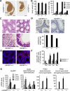
HSPBP1 is required for spermatogenesis. (A) Morphology of testes and epididymis (epi.) prepared from 8-wk-old wild-type (+/+) and HSPBP1-deficient mice (−/−). Scale bars, 2 mm. (B) Body weight and testis weight of wild-type (+/+) and transgenic mice heterozygous (+/−) and homozygous (−/−) for HSPBP1 were determined at the indicated time points after birth. (C) Histological sections of testes of 8-wk-old HSPBP1+/+ and HSPBP1/ mice. Scale bars, 50 μm (low magnification), 20 μm (high magnification). (D) Epididymides of HSPBP1/ mice are devoid of mature sperm and show reduced weight. Top, hematoxylin and eosin staining of epididymides sections from HSPBP1+/+ and HSPBP1/ mice. Scale bars, 50 μm. Bottom, epididymis weight was determined at 8 wk after birth (mean ± SEM, n ≥ 3). (E) Seminiferous tubules in HSPBP1/ mice show an increased number of apoptotic cells. TUNEL analysis of testes sections at 8 wk after birth. Scale bars, 50 μm. (F) Quantification of data obtained in E. (G) Transcript levels for HSPBP1 and stage-specific spermatogenesis markers determined in testis lysates by qRT-PCR at the indicated time points after birth. Maximum level detected in wild-type mice was set to 100% (mean ± SEM, n ≥ 3).
FIGURE 3
FIGURE 3
Ablation of HSPBP1 expression results in a late pachytene arrest of meiotic spermatocytes. (A) Immunostaining of chromosome spreads from adult testes for synaptonemal complex proteins SYCP3 (axial/lateral elements) and SYCP1 (transverse filament). (B) Graph showing the distribution of SYCP3-positive nuclei across meiotic prophase substages as determined by SYCP3/SYCP1 immunostaining (n = 300, three mice of each genotype, 100 nuclei/mouse). (C) Immunostaining of adult testis chromosome spreads for SYCP3 and γH2AX (double-stranded DNA-break marker). The sex body is indicated with an arrow. (D) Immunostaining of adult testis chromosome spreads for SYCP3 and MLH1 (late recombination marker). (E) Immunostaining of adult testis chromosome spreads for SYCP3 and RBMY (marker for meiotic sex chromosome inactivation). Scale bars, 20 μm.
FIGURE 4
FIGURE 4
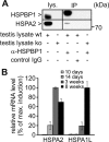
HSPBP1 and HSPA2 interact with each other in testes and are coinduced during spermatogenesis. (A) Testis lysates of wild-type (wt) and HSPBP1/ mice (ko) were prepared in RIPA buffer, and the soluble protein fraction was subjected to immunoprecipitation (IP) with an anti-HSPBP1 antibody or the same amount of control immunoglobulin G. A 40-μg amount of lysate protein was loaded as control (lys.). (B) At the indicated time points after birth, mRNA was prepared from testis, and transcript levels were determined for HSPA2 and HSPA1L by qRT-PCR. The maximum transcript level was set to 100% (see Figure 2G for HSPBP1 expression during spermatogenesis).
FIGURE 5
FIGURE 5
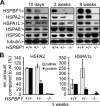
HSPBP1 ablation causes a reduction of HSPA2 and HSPA1L expression in mouse testis. (A) At the indicated time points after birth, testis lysates were prepared from wild-type (+/+) and HSPBP1-depleted mice. Chaperones and cochaperones were detected using specific antibodies. Each lane corresponds to 100 μg of soluble lysate protein. Tubulin served as loading control. (B) Protein levels of HSPA2 and HSPA1L, detected in testis lysates as shown in A, were quantified (black bars) and compared with transcript levels in testes for the two proteins determined by qRT-PCR (light gray) (mean ± SEM, n ≥ 3).
FIGURE 6
FIGURE 6
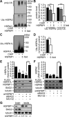
HSPBP1 interferes with the ubiquitylation of inducible HSP70 chaperones and cooperates with BAG2 in preventing the proteasomal degradation of HSPA2 and HSPA1L. (A) In vitro ubiquitylation of purified HSPA2 was performed in the presence of E1 and UBCH5b. Where indicated, purified CHIP and increasing concentrations of HSPBP1 (onefold and fivefold molar excess over added CHIP) were added to the reaction. Ubiquitin conjugates were detected by an anti-ubiquitin conjugate antibody (poly-Ub.) and anti-HSPA2 (HSPA2/Ubn-HSPA2), respectively. (B) Quantification of data obtained in A (mean ± SEM, n = 3; *p < 0.05, **p < 0.001, ***p < 0.0001). (C) In vitro ubiquitylation of purified HSPA1L was performed in the presence of E1 and UBCH5b. Where indicated, purified CHIP and HSPBP1 (at a fivefold molar excess over added CHIP) were added to the reaction. Ubiquitin conjugates were detected by an anti-HSP70 antibody. (D) Quantification of data obtained in C (mean ± SEM, n = 3; **p < 0.001). (E) Analysis of HSPA1L stability in HeLa cells. FLAG-HSPA1L–expressing cells were transfected with siRNA targeting HSPBP1 and BAG2 or a control siRNA (–) as indicated. MG132 (20 μM, 16 h) was added when indicated to verify proteasomal degradation of FLAG-HSPA1L. Cells were harvested and lysed after 72 h and analyzed by SDS–PAGE and immunoblotting to quantify protein levels of FLAG-HSPA1L (mean ± SEM, n = 3; *p < 0.05, **p < 0.001). (F) Analysis of HSPA2 stability in mouse embryonic fibroblasts. FLAG-HSPA2–expressing cells were transfected with siRNA targeting HSPBP1 and BAG2 or a control siRNA (–) as indicated. MG132 (20 μM, 16 h) was added when indicated to verify proteasomal degradation. Cells were harvested and lysed after 48 h and analyzed by SDS–PAGE and immunoblotting to quantify protein levels (mean ± SEM, n = 3; *p < 0.05, **p < 0.001). (G) Detection of HSPBP1 and BAG2 in lysates of brain and testis from 3-wk-old wild-type (+/+), HSPBP1 heterozygous (+/−), and homozygous (−/−) knockout mice. Each lane corresponds to 100 μg of soluble lysate protein. The cochaperones were detected with specific antibodies. Tubulin served as loading control.
FIGURE 7
FIGURE 7
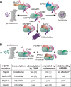
Role of HSPBP1 and BAG2 as regulators of CHIP-mediated degradation. (A) Schematic presentation of the HSP70/cochaperone network that regulates CHIP-mediated protein degradation. CHIP binds to the carboxy terminus of HSP70 and ubiquitylates the chaperone and bound client proteins in conjunction with ubiquitin-conjugating enzymes of the UBCH4/5 family. Targeting to the proteasome is assisted by the cochaperone BAG1, whereas binding of BAG3 to the chaperone/CHIP complex induces the lysosomal degradation of the client. HSPBP1 and BAG2 inhibit the ubiquitin ligase activity of CHIP in the assembled chaperone complex and thereby facilitate the folding of the client. (B) Schematic presentation of the CHIP/HSPBP1/BAG2 cochaperone network that balances the expression of inducible HSP70 chaperones at the posttranslational level. CHIP is able to mediate the ubiquitylation of inducible HSP70s and induces their proteasomal degradation. Association of HSPBP1 or BAG2 with the HSP70/CHIP complex results in an inhibition of CHIP activity, leading to stabilization of inducible HSP70s. *Present study; (1) Jiang et al. (2001), (2) Qian et al. (2006), (3) Sasaki et al. (2008).

References

    1. Akerfelt M, Vihervaara A, Laiho A, Conter A, Christians ES, Sistonen L, Henriksson E. Heat shock transcription factor 1 localizes to sex chromatin during meiotic repression. J Biol Chem. 2010;285:34469–34476. - PMC - PubMed
    1. Alberti S, Bohse K, Arndt V, Schmitz A, Höhfeld J. The cochaperone HspBP1 inhibits the CHIP ubiquitin ligase and stimulates the maturation of the cystic fibrosis transmembrane conductance regulator. Mol Biol Cell. 2004;15:4003–4010. - PMC - PubMed
    1. Alberti S, Demand J, Esser C, Emmerich N, Schild H, Höhfeld J. Ubiquitylation of BAG-1 suggests a novel regulatory mechanism during the sorting of chaperone substrates to the proteasome. J Biol Chem. 2002;277:45920–45927. - PubMed
    1. Alekseev OM, Richardson RT, O'Rand MG. Linker histones stimulate HSPA2 ATPase activity through NASP binding and inhibit CDC2/cyclin B1 complex formation during meiosis in the mouse. Biol Reprod. 2009;81:739–748. - PMC - PubMed
    1. Allen JW, Dix DJ, Collins BW, Merrick BA, He C, Selkirk JK, Poorman-Allen P, Dresser ME, Eddy EM. HSP70–2 is part of the synaptonemal complex in mouse and hamster spermatocytes. Chromosoma. 1996;104:414–421. - PubMed

Publication types

Substances

LinkOut - more resources