. 2014 Jun 4;34(23):8032-42.
doi: 10.1523/JNEUROSCI.0236-14.2014.

The substantia nigra conveys target-dependent excitatory and inhibitory outputs from the basal ganglia to the thalamus

Affiliations

The substantia nigra conveys target-dependent excitatory and inhibitory outputs from the basal ganglia to the thalamus

Miklos Antal et al. J Neurosci. .

Abstract

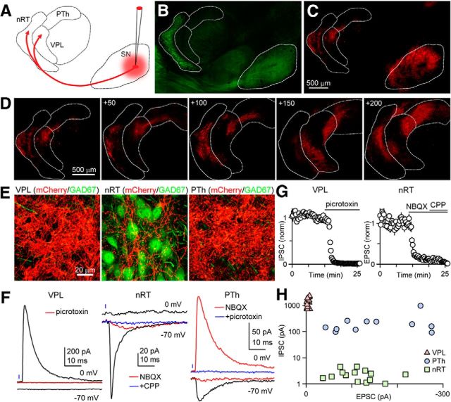
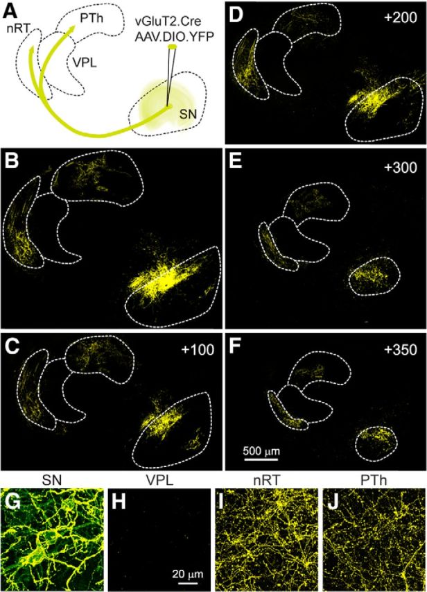
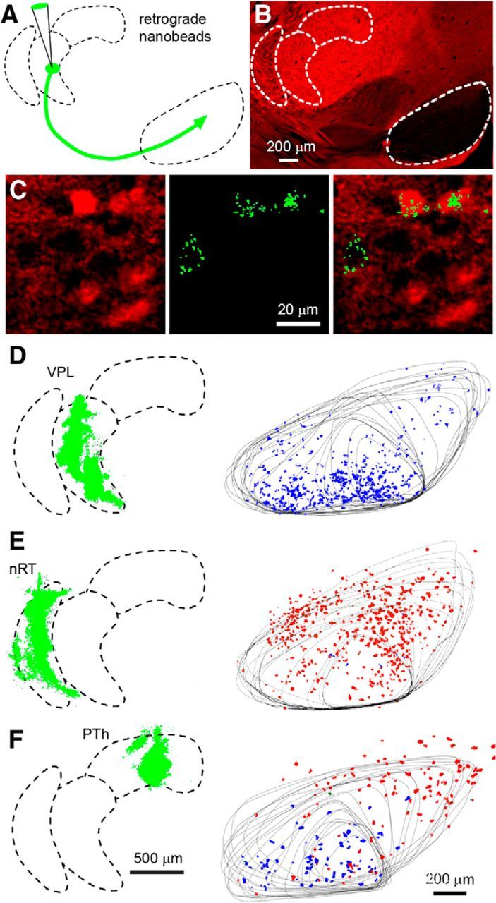
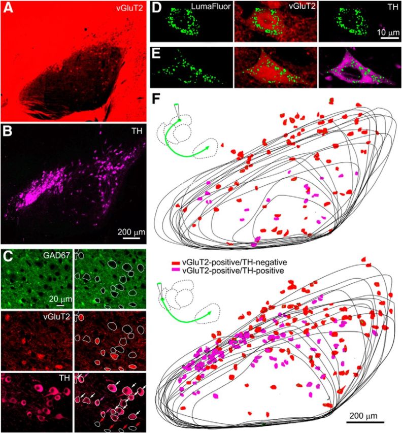
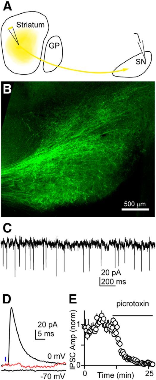
The basal ganglia (BG), which influence cortical activity via the thalamus, play a major role in motor activity, learning and memory, sensory processing, and many aspects of behavior. The substantia nigra (SN) consists of GABAergic neurons of the pars reticulata that inhibit thalamic neurons and provide the primary output of the BG, and dopaminergic neurons of the pars compacta that modulate thalamic excitability. Little is known about the functional properties of the SN→thalamus synapses, and anatomical characterization has been controversial. Here we use a combination of anatomical, electrophysiological, genetic, and optogenetic approaches to re-examine these synaptic connections in mice. We find that neurons in the SN inhibit neurons in the ventroposterolateral nucleus of the thalamus via GABAergic synapses, excite neurons in the thalamic nucleus reticularis, and both excite and inhibit neurons within the posterior nucleus group. Glutamatergic SN neurons express the vesicular glutamate receptor transporter vGluT2 and receive inhibitory synapses from striatal neurons, and many also express tyrosine hydroxylase, a marker of dopaminergic neurons. Thus, in addition to providing inhibitory outputs, which is consistent with the canonical circuit, the SN provides glutamatergic outputs that differentially target thalamic nuclei. This suggests that an increase in the activity of glutamatergic neurons in the SN allows the BG to directly excite neurons in specific thalamic nuclei. Elucidating an excitatory connection between the BG and the thalamus provides new insights into how the BG regulate thalamic activity, and has important implications for understanding BG function in health and disease.

Keywords: circuitry; substantia nigra; vGluT2.


Figures

Figure 1.
Figure 1.
The SN provides both excitatory and inhibitory connections to the thalamic nuclei in a target-dependent manner. A, Schematic showing AAV injection to express ChR2-mCherry in SN neurons cells. Thalamic regions are labeled nRT, VPL, and PTh. B, The injection was performed in a GAD67-GFP mouse, in which inhibitory neurons are labeled in green. The SN and the nRT, which contain primarily GABAergic neurons, are readily identified. C, mCherry fluorescence shows prominent expression in SN cell bodies in the SN and fibers in the thalamus. D, Fiber labeling within the thalamus is shown for multiple consecutive sections, with the first panel corresponding to plate 11. Coordinates indicate steps, in micrometers, toward midline. E, High-power images of red and green fluorescence show the labeled fibers (mCherry) and GABAergic neurons (green in GAD67-GFP mice) within different thalamic regions. F, Representative light-evoked responses are shown for the different nuclei. Glutamate-mediated currents were recorded at −70 mV, while GABAA-mediated responses were studied at 0 mV. In the VPL, no excitatory response was observed, but an inhibitory current at 0 mV was eliminated with a GABAAR antagonist (red, picrotoxin). In the nRT, no excitatory component was observed, but an excitatory component was observed that greatly attenuated to an AMPAR antagonist (red, NBQX) and was eliminated by the subsequent addition of an NMDAR antagonist (blue, CPP). In the PTh, an excitatory component was observed (black, −70 mV) that was almost eliminated by NBQX (red). A light-evoked response was also observed at 0 mV that was unaffected by NBQX (red), indicating a direct, monosynaptic inhibitory response, which was eliminated by picrotoxin. G, Summary of pharmacological characterization of light-evoked responses using a GABAAR antagonist (picrotoxin), an AMPAR antagonist (NBQX), and an NMDAR antagonist (R-CPP). H, Summary of the amplitudes of light-evoked IPSCs and EPSCs in different thalamic regions.
Figure 2.
Figure 2.
GABAergic and glutamatergic neurons in the substantia nigra form two non-overlapping populations as identified in GAD67::GFP/vGluT2::tdTomato transgenic mice. A, Low-magnification view of widespread green GFP epifluorescent signal due to the abundant presence of GABAergic somata and processes within the SN. This sagittal slice was taken from a mouse line expressing both tdTomato and GFP genes in vGluT2-positive and GAD67-positive cell populations, respectively (vGluT2-Cre::tdTomato/GAD67::GFP). B, A sparser and more peripheral but substantial red tdTomato epifluorescent signal is exhibited in the same slice by vGluT2-positive neurons within the SN. C, Confocal merged image of green and red fluorescence demonstrating the mutual exclusivity of the GABAergic and glutamatergic neuron populations. D, vGluT2-positive neurons were identified by red fluorescence. Representative example of the schematization process during which the glutamatergic cell bodies were colored black and the background removed. Neurons expressing both vGluT2 (red) and GAD67 (green) were not observed. E–J, The location of vGluT2-positive neurons in SN, identified as in D, is shown for six sections from the same hemisphere. The representative schematized images of SN slice preparations were taken at lateral (A), intermediate (F–H), and medial (I, J) sagittal planes.
Figure 3.
Figure 3.
Glutamatergic neurons in the substantia nigra send projections to the nRT and the PTh, but not the VPL. A, Schematic showing the conditional viral expression and the injection site in a vGluT2-Cre mouse. B–F, Successive sections showing the YFP labeling in the SN and thalamus, with the first panel corresponding to plate 13. Coordinates indicate steps, in micrometers, toward the midline. G–J, High-power images of the labeling observed in the SN and in different thalamic regions.
Figure 4.
Figure 4.
Retrograde labeling of SN neurons following nanobead injection into thalamic nuclei. A, Schematic showing the injection of nanobeads into the thalamus of a vGluT2-Cre::tdTomato mouse. B, The injections were performed in vGluT2-Cre::tdTomato mice in which vGluT2-positive neurons are labeled in red. Distinct regions exhibiting differential vGluT2 density are readily identified. C, High-power image in the SN in which neurons that express vGluT2 are red (left), retrogradely labeled neurons show characteristic punctate green fluorescence (middle), and the merged image shows that, following this injection into the PTh, one non-glutamatergic and two glutamatergic neurons were labeled. D, Shows an experiment in which beads were injected into the VPL, with the injection site shown as a Z-stack determined from 17 sections (left, green). Twenty-one sections were used to determine the location of bead-labeled neurons in the SN, with the outline of the SN shown for all of the sections, and bead-labeled neurons were color coded, with red indicating vGluT2-positive neurons and blue indicating vGluT2-negative neurons. E, Example of retrograde labeling following bead injection into the nRT. F, Example of retrograde labeling following bead injection in the PTh.
Figure 5.
Figure 5.
Many of the vGluT2-positive neurons in the SN that project to the nRT and the PTh are dopaminergic. A, Fluorescence in a vGluT2-tdTomato mouse. B, An antibody to TH (purple) was used to identify dopaminergic neurons in the same preparation as in A. C, Fluorescence is shown for a GAD67-GFP::vGluT2-TdTomato mouse stained with a TH antibody. On the right, cells are circled and indicated by arrows (bottom right): vGluT2-positive/TH-negative (red arrows), vGluT2-postitive/TH-positive (purple arrows), and vGluT2-negative/TH-positive (white arrows). D–F, Experiments were performed in which fluorescent nanobeads were injected into the thalamus of vGluT2::tdTomato mice stained with a TH antibody. High-power images of two cells labeled with fluorescent nanobeads reveal a vGluT2-positive/TH-negative cell (D) and a vGluT2-positive/TH-positive cell (E). F, The location and properties of neurons labeled following thalamic injections is shown.
Figure 6.
Figure 6.
Glutamatergic neurons in the substantia nigra fire spontaneously and receive input from the striatum. A, Schematic showing injection of an AAV construct to express ChR2-YFP in striatal neurons in a vGluT2-Cre::tdTomato mouse. B, YFP labeling of striatal fibers in the SN. C, Example showing spontaneous activity of a vGuT2-positive neuron recorded with an on-cell electrode. The neuron was identified by red fluorescence in a vGuT2-Cre::tdTomato mouse. D, Example showing whole-cell recording from a vGluT2-positive cell. The IPSC recorded at 0 mV was eliminated by pictrotoxin (red). E, Summary of recordings from 10 cells showing the blockade of the IPSC with a GABAAR antagonist.

References

    1. Adamantidis AR, Tsai HC, Boutrel B, Zhang F, Stuber GD, Budygin EA, Touriño C, Bonci A, Deisseroth K, de Lecea L. Optogenetic interrogation of dopaminergic modulation of the multiple phases of reward-seeking behavior. J Neurosci. 2011;31:10829–10835. doi: 10.1523/JNEUROSCI.2246-11.2011. - DOI - PMC - PubMed
    1. Aggleton JP, Neave N, Nagle S, Sahgal A. A comparison of the effects of medial prefrontal, cingulate cortex, and cingulum bundle lesions on tests of spatial memory: evidence of a double dissociation between frontal and cingulum bundle contributions. J Neurosci. 1995;15:7270–7281. - PMC - PubMed
    1. Aggleton JP, Hunt PR, Nagle S, Neave N. The effects of selective lesions within the anterior thalamic nuclei on spatial memory in the rat. Behav Brain Res. 1996;81:189–198. doi: 10.1016/S0166-4328(96)89080-2. - DOI - PubMed
    1. Alexander GE, Crutcher MD. Functional architecture of basal ganglia circuits: neural substrates of parallel processing. Trends Neurosci. 1990;13:266–271. doi: 10.1016/0166-2236(90)90107-L. - DOI - PubMed
    1. Alexander GE, Crutcher MD, DeLong MR. Basal ganglia-thalamocortical circuits: parallel substrates for motor, oculomotor, “prefrontal” and “limbic” functions. In: Uylings HBM, editor. The prefrontal cortex: its structure, function and pathology; Progress in Brain Research. New York: Elsevier; 1991. pp. 119–146. - PubMed

Publication types

MeSH terms

Substances