. 2011 Jun;45(2):117-24.
doi: 10.1007/s13139-011-0081-0. Epub 2011 Apr 20.

Prognostic Value of Primary Tumor Uptake on F-18 FDG PET/CT in Patients with Invasive Ductal Breast Cancer

Affiliations

Prognostic Value of Primary Tumor Uptake on F-18 FDG PET/CT in Patients with Invasive Ductal Breast Cancer

Bong-Il Song et al. Nucl Med Mol Imaging. 2011 Jun.

Abstract

Purpose: To determine the prognostic implications of pretreatment F-18 FDG PET/CT in patients with invasive ductal breast cancer (IDC), we evaluated the relationship between FDG uptake of the primary tumor and known prognostic parameters of breast cancer. Prognostic significance of tumoral FDG uptake for the prediction of progression-free survival (PFS) was also assessed.

Materials and methods: Fifty-five female patients with IDC who underwent pretreatment F-18 FDG PET/CT were enrolled. The maximum standardized uptake value of the primary tumor (pSUVmax) was compared with clinicopathological parameters including tumor size, grade, estrogen receptor (ER), progesterone receptor (PR), human epidermal growth factor receptor2 (HER2), axillary lymph node (LN) metastasis, and stage. The prognostic value of pSUVmax for PFS was assessed using the Kaplan-Meier method.

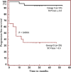
Results: A high pSUVmax was significantly related to a higher stage of tumor size (P < 0.05), grade (P < 0.001), and stage (P < 0.001). pSUVmax was significantly higher in ER-negative tumors (P < 0.001), PR-negative tumors (P < 0.001), and positive LN metastasis (P < 0.01), but not different according to HER2 status. pSUVmax was significantly higher in patients with progression compared to patients who were disease-free (10.6 ± 5.1 vs. 4.7 ± 3.5, P < 0.001). A receiver-operating characteristic curve demonstrated a pSUVmax of 6.6 to be the optimal cutoff for predicting PFS (sensitivity; 86.7%, specificity; 82.5%). The patients with a high pSUVmax (more than 6.6) had significantly shorter PFS compared to patients with a low pSUVmax (P < 0.0001).

Conclusions: pSUVmax on pretreatment F-18 FDG PET/CT could be used as a good surrogate marker for the prediction of progression in patients with IDC.

Keywords: F-18 FDG PET/CT; Invasive ductal breast cancer; Prognosis; SUVmax.


Figures

Fig. 1
Fig. 1
pSUVmax differences according to disease group. Mann-Whitney U-test reveals a significant difference between the disease-free group and progression group (P < 0.001). Mean values of pSUVmax (4.7 in the disease-free group and 10.6 in the progression group) are indicated with purple boxes. The error bars represent the 95% confidence interval for mean
Fig. 2
Fig. 2
Optimal cutoff of pSUVmax for predicting progression-free survival
Fig. 3
Fig. 3
Progression-free survival according to the pSUVmax
Fig. 4
Fig. 4
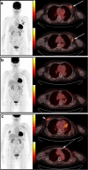
A 71-year-old female patient diagnosed with invasive ductal breast cancer (pSUVmax 7.0, tumor size 3.6 cm, ER-, PR-, HER2+). a F-18 FDG PET/CT before cancer treatment. In the pre-treatment, a focal hypermetabolic lesion in the left breast and enlarged LNs with focal FDG uptake in the left axillary area were shown, which were histologically confirmed as malignancy in both lesions (long arrow). b In the follow-up F-18 FDG PET/CT taken after neoadjuvant chemotherapy and operation, there was no evidence of residual malignancy. c But 6 months later, a newly found hypermetabolic lesion in the right breast on the 2nd follow-up F-18 FDG PET/CT was shown and histologically confirmed as metastasis (short arrow). In addition, FDG uptake in the left internal mammary LN was shown (long dotted arrow)
Fig. 5
Fig. 5
A 43-year-old female patient diagnosed with invasive ductal breast cancer (pSUVmax 2.9, tumor size 2.2 cm, ER+, PR+, HER2+). In initial F-18 FDG PET/CT, a mildly hypermetabolic lesion was shown (pSUVmax is lesser than 6.6) and histologically confirmed as malignancy in the right breast (long arrow). In the follow-up F-18 FDG PET/CT taken after the operation, chemotherapy, and hormonal therapy, there was no evidence of residual or recurrent malignancy

Similar articles

Cited by

References

    1. Desantis C, Center MM, Siegel R, Jemal A. American Cancer Society. Breast Cancer Facts & Figures 2009–2010 Atlanta: American Cancer Society, Inc. http://www.cancer.org/acs/groups/content/@nho/documents/document/f861009...
    1. Lavayssière R, Cabée AE, Filmont JE. Positron emission tomography (PET) and breast cancer in clinical practice. Eur J Radiol. 2009;69:50–58. doi: 10.1016/j.ejrad.2008.07.039. - DOI - PubMed
    1. Geus-Oei LF, Oyen WJ. Predictive and prognostic value of FDG-PET. Cancer Imaging. 2008;8:70–80. doi: 10.1102/1470-7330.2008.0010. - DOI - PMC - PubMed
    1. Allal AS, Slosman DO, Kebdani T, Allaoua M, LehmannW DP. Prediction of outcome in head-and-neck cancer patients using the standardized uptake value of 2-[18F]fluoro-2-deoxy-D-glucose. Int J Radiat Oncol Biol Phys. 2004;59:1295–1300. doi: 10.1016/j.ijrobp.2003.12.039. - DOI - PubMed
    1. Downey RJ, Akhurst T, Gonen M, Vincent A, Bains MS, Larson S, et al. Preoperative F-18 fluorodeoxyglucose-positron emission tomography maximal standardized uptake value predicts survival after lung cancer resection. J Clin Oncol. 2004;22:3255–3260. doi: 10.1200/JCO.2004.11.109. - DOI - PubMed

LinkOut - more resources