

Intratumoral Heterogeneous F-18 Fluorodeoxyglucose Uptake Corresponds with Glucose Transporter-1 and Ki-67 Expression in a Case of Krukenberg Tumor: Localization of Intratumoral Hypermetabolic Focus by Fused PET/MR Image
- PMID: 24899993
- PMCID: PMC4043022
- DOI: 10.1007/s13139-010-0071-7
Intratumoral Heterogeneous F-18 Fluorodeoxyglucose Uptake Corresponds with Glucose Transporter-1 and Ki-67 Expression in a Case of Krukenberg Tumor: Localization of Intratumoral Hypermetabolic Focus by Fused PET/MR Image
Abstract
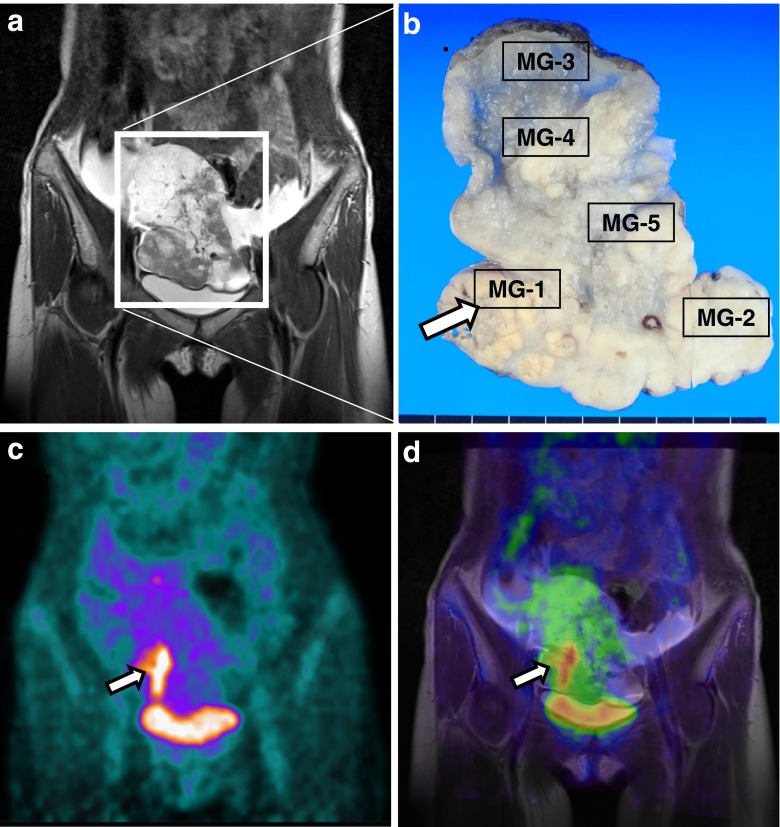
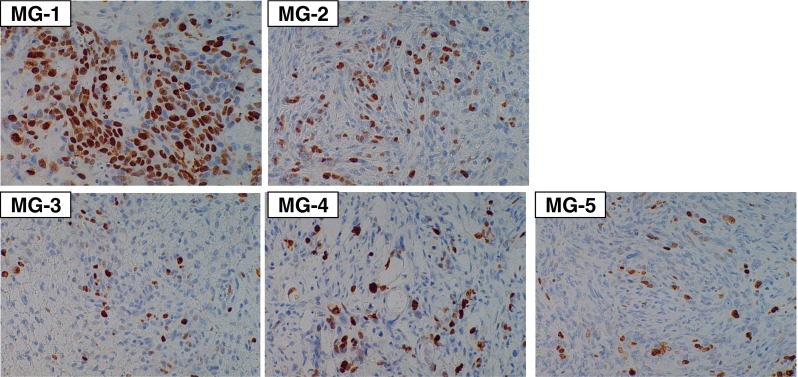
The expression of glucose transporters (Glut-1, Glut-3), hexokinase-II, and Ki-67 has been proposed to explain intratumoral heterogeneous F-18 fluorodeoxyglucose (FDG) uptake. We report a case of Krukenberg tumor with intratumoral heterogeneous FDG uptake which corresponded well with the expression levels of Glut-1 and ki-67. Fused positron emission tomography (PET)/magnetic resonance (MR) imaging was helpful for localizing the metabolically active area in the tumor specimen. This report elucidates the relationship between the intratumoral heterogeneous FDG uptake and biologic heterogeneity, and shows the usefulness of PET/MR in research on intratumoral heterogeneity.
Keywords: FDG; Glut-1; Ki-67; Krukenberg tumor; PET/CT; PET/MRI.
Figures


Similar articles
-
Biologic correlates of intratumoral heterogeneity in 18F-FDG distribution with regional expression of glucose transporters and hexokinase-II in experimental tumor.J Nucl Med. 2005 Apr;46(4):675-82. J Nucl Med. 2005. PMID: 15809491
-
Biologic correlation between glucose transporters, hexokinase-II, Ki-67 and FDG uptake in malignant melanoma.Nucl Med Biol. 2012 Nov;39(8):1167-72. doi: 10.1016/j.nucmedbio.2012.07.003. Epub 2012 Aug 15. Nucl Med Biol. 2012. PMID: 22901702
-
Biological characteristics of intratumoral [F-18]‑fluoromisonidazole distribution in a rodent model of glioma.Int J Oncol. 2013 Mar;42(3):823-30. doi: 10.3892/ijo.2013.1781. Epub 2013 Jan 18. Int J Oncol. 2013. PMID: 23338175 Free PMC article.
-
2-[18F]-2-deoxy-D-glucose (FDG) uptake in human tumor cells is related to the expression of GLUT-1 and hexokinase II.Acta Radiol. 2008 Dec;49(10):1145-53. doi: 10.1080/02841850802482486. Acta Radiol. 2008. PMID: 18979289
-
18F-fluorodeoxyglucose positron emission tomography/computed tomography and the relationship between fluorodeoxyglucose uptake and the expression of hypoxia-inducible factor-1α, glucose transporter-1 and vascular endothelial growth factor in thymic epithelial tumours.Eur J Cardiothorac Surg. 2013 Aug;44(2):e105-12. doi: 10.1093/ejcts/ezt263. Epub 2013 May 14. Eur J Cardiothorac Surg. 2013. PMID: 23674658
References
-
- Hockel M, Schlenger K, Aral B, Mitze M, Schaffer U, Vaupel P. Association between tumor hypoxia and malignant progression in advanced cancer of the uterine cervix. Cancer Res. 1996;56:4509–4515. - PubMed
LinkOut - more resources
Full Text Sources
Miscellaneous