

Necrotizing Cervical Lymphadenitis Caused by Mycobacterium simiae in an HIV-Positive Patient: Imaging with (18)F-FDG PET/CT
- PMID: 24900008
- PMCID: PMC4043006
- DOI: 10.1007/s13139-011-0088-6
Necrotizing Cervical Lymphadenitis Caused by Mycobacterium simiae in an HIV-Positive Patient: Imaging with (18)F-FDG PET/CT
Abstract
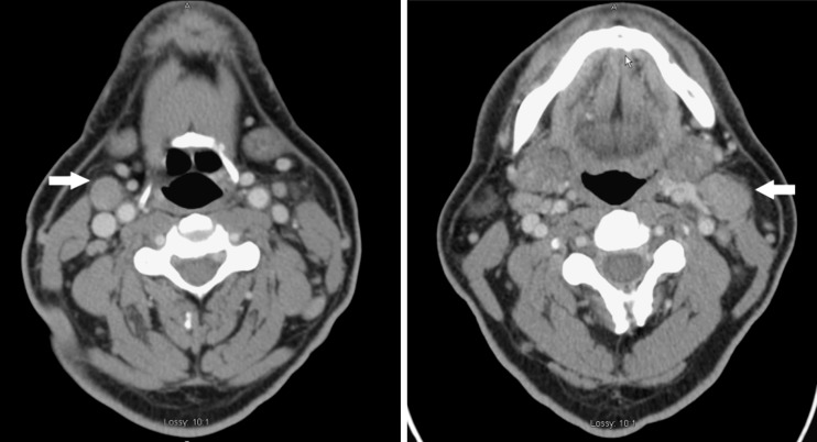
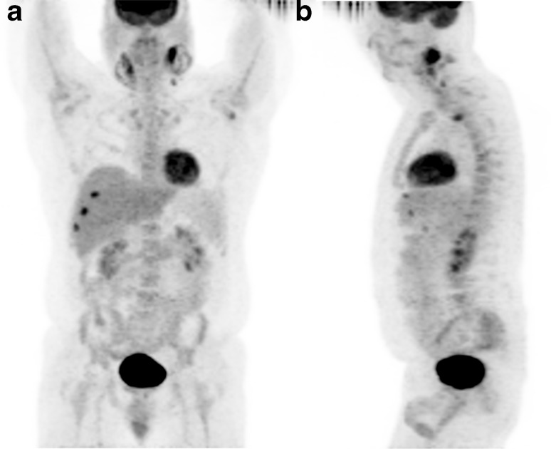
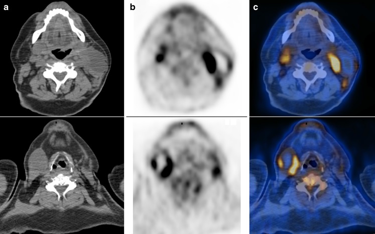
Mycobacterium simiae (M. simiae) is an opportunistic pathogen rarely associated with human disease, although in recent years M. simiae has been detected with increasing frequency in human immunodeficiency virus (HIV)-infected patients, usually causing disseminated infection with fever, diarrhea and weight loss. We report the case of an HIV-positive man, who was referred for an (18)F-FDG PET/CT to evaluate a solitary pulmonary nodule. The PET/CT showed incidental large necrotic cervical lymph nodes, compatible with necrotizing cervical lymphadenitis. Biopsy and culture of one of the affected lymph nodes were positive for M. simiae. We present the first report of (18)F-FDG PET/CT imaging of an infectious process caused by M. simiae in humans.
Keywords: Fluorodeoxyglucose; HIV; Mycobacterium simiae; Necrotizing cervical lymphadenitis; PET/CT.
Figures
Similar articles
-
Mycobacterium simiae cervical lymphadenitis.Pediatr Infect Dis J. 2007 Apr;26(4):362-3. doi: 10.1097/01.inf.0000258614.98241.4e. Pediatr Infect Dis J. 2007. PMID: 17414407
-
Successful treatment of disseminated Mycobacterium simiae infection in AIDS patients.Scand J Infect Dis. 1998;30(2):143-6. doi: 10.1080/003655498750003528. Scand J Infect Dis. 1998. PMID: 9730300
-
Immune reconstitution inflammatory syndrome due to Mycobacterium avium complex successfully followed up using 18 F-fluorodeoxyglucose positron emission tomography-computed tomography in a patient with human immunodeficiency virus infection: A case report.BMC Med Imaging. 2015 Jul 18;15:24. doi: 10.1186/s12880-015-0063-2. BMC Med Imaging. 2015. PMID: 26187282 Free PMC article.
-
Positron emission tomography/computed tomography hypermetabolism of Kikuchi-Fujimoto disease mimicking malignant lymphoma: a case report and literature review.J Int Med Res. 2021 Jul;49(7):3000605211032859. doi: 10.1177/03000605211032859. J Int Med Res. 2021. PMID: 34334002 Free PMC article. Review.
-
About two cases of Mycobacterium simiae infection in AIDS: review of the pathogenicity.Acta Clin Belg. 1998 Jun;53(3):206-12. Acta Clin Belg. 1998. PMID: 9701858 Review.
Cited by
-
Kikuchi Disease Mimicking Metastatic Lymphadenopathy on (18)F-FDG PET/CT in Patients with Breast Cancer.Nucl Med Mol Imaging. 2015 Jun;49(2):167-8. doi: 10.1007/s13139-015-0318-4. Epub 2015 Jan 23. Nucl Med Mol Imaging. 2015. PMID: 26082813 Free PMC article. No abstract available.
-
Mycobacterium simiae: Harmless colonizer or deadly pathogen?PLoS Pathog. 2020 Apr 30;16(4):e1008418. doi: 10.1371/journal.ppat.1008418. eCollection 2020 Apr. PLoS Pathog. 2020. PMID: 32353074 Free PMC article. Review. No abstract available.
References
LinkOut - more resources
Full Text Sources