Case Reports
. 2012 Sep;46(3):218-22.
doi: 10.1007/s13139-012-0150-z. Epub 2012 Jun 27.

Hibernoma: Intense Uptake on F18-FDG PET/CT

Affiliations
Case Reports

Hibernoma: Intense Uptake on F18-FDG PET/CT

Jong Deok Kim et al. Nucl Med Mol Imaging. 2012 Sep.

Abstract

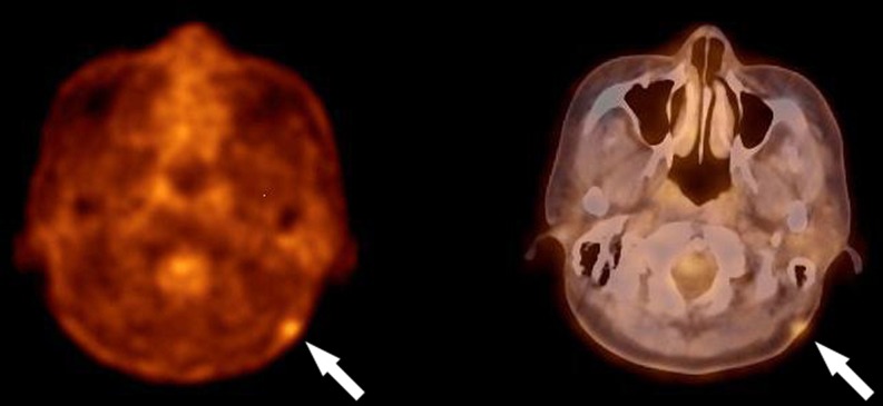
Hibernoma is a rare benign but metabolically active tumor of brown fat origin, that can have cross-sectional imaging characteristics similar to those of other fat-containing tumors, such as lipomas and liposarcomas, and its presence can lead to false-positive interpretation by exhibiting increased F18-fluorodeoxyglucose (F18-FDG) activity. A 46-year-old woman was diagnosed with dermatofibrosarcoma protuberans underwent F18-FDG positron emission tomography/computed tomography (PET/CT) for detecting recurrence after excision. F18-FDG PET/CT showed incidental intense uptake in the back in addition to increased F18-FDG uptake at the previous lesion site. To our knowledge, this is the first case report of intense F18-FDG uptake in hibernoma in Korea.

Keywords: Dermatofibrosarcoma protuberance; Hibernoma; PET/CT.


Figures

Fig. 1
Fig. 1
Transaxial F18-FDG PET/CT images show a recurrent dermatofibrosarcoma protuberans in the left retromastoid area with SUVmax of 2.75
Fig. 2
Fig. 2
Transaxial (a, b), sagittal (c, d) and coronal (e, f) F18-FDG PET/CT images show a 2 × 4 × 4.5-cm sized hypermetabolic mass in the subcutaneous layer of the left back at T9-T10 level with SUVmax of 12.83 in 1 h
Fig. 3
Fig. 3
Contrast-enhanced MRI images show the left back mass in subcutaneous layer at T9-T10 level with heterogenous T1 and T2 hyperintensity (hypointense to adjacent subcutaneous fat) (a, b) and intense enhancement with a linear hyperintensity entering the mass (c, d). The mass is not suppressed on fat-saturation image (e)
Fig. 4
Fig. 4
Microscopic view of the tumor shows round or polygonal multivacuolated cells and mature adipose cells, and many small vessels

References

    1. Little BP, Fintlemann FJ, Kenudson MM, Lanuti M, Shepard JO, Digumarthy SR. Intrathoracic hibernoma; a case with multimodality imaging correlation. J Thorac Imaging. 2011;26:W20–W22. doi: 10.1097/RTI.0b013e3181e35acd. - DOI - PubMed
    1. Himms-Hagen J. Thermogenesis in brown adipose tissue as an energy buffer. N Engl J Med. 1984;311:1549–1558. doi: 10.1056/NEJM198412133112407. - DOI - PubMed
    1. Yeung HW, Grewal RK, Gonen M, Schoder H, Larson S. Patterns of F18-FDG uptake in adipose tissue and muscle: a potential sourse of false-positives for PET. J Nucl Med. 2003;44:1789–1796. - PubMed
    1. Furlong MA, Famburg-Smith JC, Miettinen M. The morphologic spectrum of hibernoma: a clinocopathologic study of 170 cases. Am J Surg Pathol. 2001;25(6):809–814. doi: 10.1097/00000478-200106000-00014. - DOI - PubMed
    1. Bancroft L, Kransdorf M, Peterson J, O'Connor M. Benign fatty tumors: classification, clinical course, imaging appearance and treatment. Skelet Radiol. 2006;35:719–733. doi: 10.1007/s00256-006-0189-y. - DOI - PubMed

Publication types

LinkOut - more resources