

Uterine Epithelioid Angiosarcoma on F-18 FDG PET/CT
- PMID: 24900095
- PMCID: PMC4041971
- DOI: 10.1007/s13139-013-0191-y
Uterine Epithelioid Angiosarcoma on F-18 FDG PET/CT
Abstract
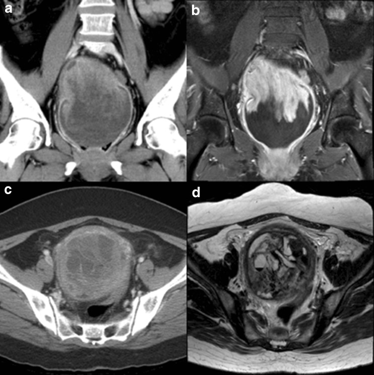
Uterine epithelioid angiosarcoma can have conventional imaging characteristics similar to those of other uterine tumors, such as leiomyoma, leiomyosarcomas or hemangioendothelioma. Uterine epithelioid angiosarcoma exhibiting increased fluorine-18 fluorodeoxyglucose (F-18 FDG) activity can be misdiagnosed. A 61-year-old woman who was diagnosed with uterine epithelioid angiosarcoma underwent F-18 FDG positron emission tomography/computed tomography (PET/CT) as a part of the pretreatment work up for surgery. F-18 FDG PET/CT showed an intense F-18 FDG uptake in the uterus in addition to increased F-18 FDG uptake at the paraaortic and aortocaval lymph nodes. To our knowledge, this is the first case report of intense F-18 FDG uptake in uterine epithelioid angiosarcoma in Korea.
Keywords: F-18 FDG; PET/CT; Uterine epithelioid angiosarcoma.
Figures

References
-
- Abrahamson TG, Stone MS, Piette WW. Cutaneous angiosarcoma. Adv Dermatol. 2001;17:279–299. - PubMed
-
- Fletcher CDM. Diagnostic histopathology of tumors. 3. Philadelphia: Elsevier Limited; 2007. pp. 66–67.
LinkOut - more resources
Full Text Sources
Other Literature Sources