

Review
doi: 10.1007/s12410-012-9160-6.
Multimodality Imaging in Congenital Heart Disease: an Update
Affiliations
- PMID: 24900167
- PMCID: PMC4032470
- DOI: 10.1007/s12410-012-9160-6
Item in Clipboard
Review
Multimodality Imaging in Congenital Heart Disease: an Update
Curr Cardiovasc Imaging Rep.
2012.
Abstract
The increasing number of survivors of congenital heart disease (CHD) has been paralleled by advancement of imaging modalities used for the ongoing assessment of these patients. There has been a large body of literature describing new approaches to non-invasive assessment of CHD. We will review new applications of well established as well as novel techniques for the management and understanding of CHD.
Keywords: Angiography; Computed tomography; Congenital heart disease; Echocardiography; Magnetic resonance imaging; Multimodality imaging.
Figures

3-dimensional echocardiography of a patient with hypoplastic left heart syndrome by transthoracic approach (a) and in a normal subject by transesophageal approach (b). In each box, images include the apical view (top left), 2-chamber view (top right), axial view (bottom left), and pyramidal full volume of the heart (bottom right)
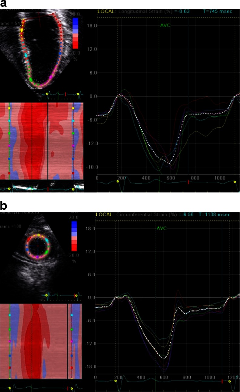
Longitudinal strain obtained in the 4-chamber view of a normal patient (a); note uniformity of strain curves from the different segments of the myocardium. Circumferential strain curve of the left ventricle in short axis derived normal patient (b)
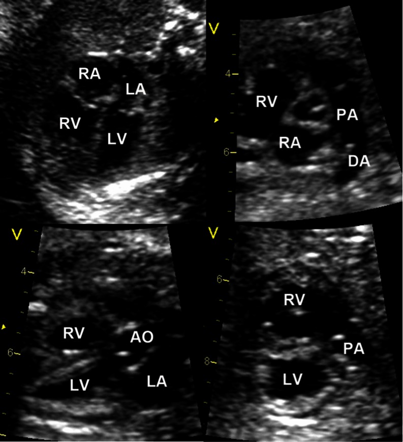
Fetal echocardiogram during the second trimester can clearly show cardiac anatomy and ventricular size. This image depicts a normal 4-chamber view of the heart (top left), short axis (top right), left ventricular outflow tract (bottom left), and right ventricular outflow tract (bottom right)

MRI tissue tagging sequence in a normal patient depicting regions of myocardium in systole (left) and diastole (right)
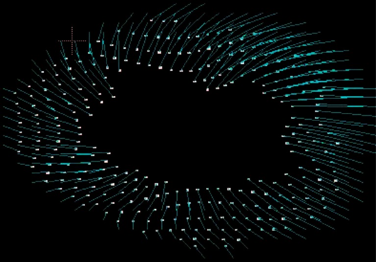
DENSE imaging of the left ventricle. Note the vectors depicting outward movement during diastole

4D flow imaging of a tortuous aorta (a) with velocity change through the region of the distorted isthmus, as shown by the color change of flow. 4D depiction of vortex with a normal left ventricle with the highest velocity seen in the center of the chamber (b)

Fetal cardiac MRI by black blood (top left) and fast SSFP through the axial plane (bottom left) of the heart, and the entire fetus (right). Note delineation of the ventricular chamber size in the 4-chamber view
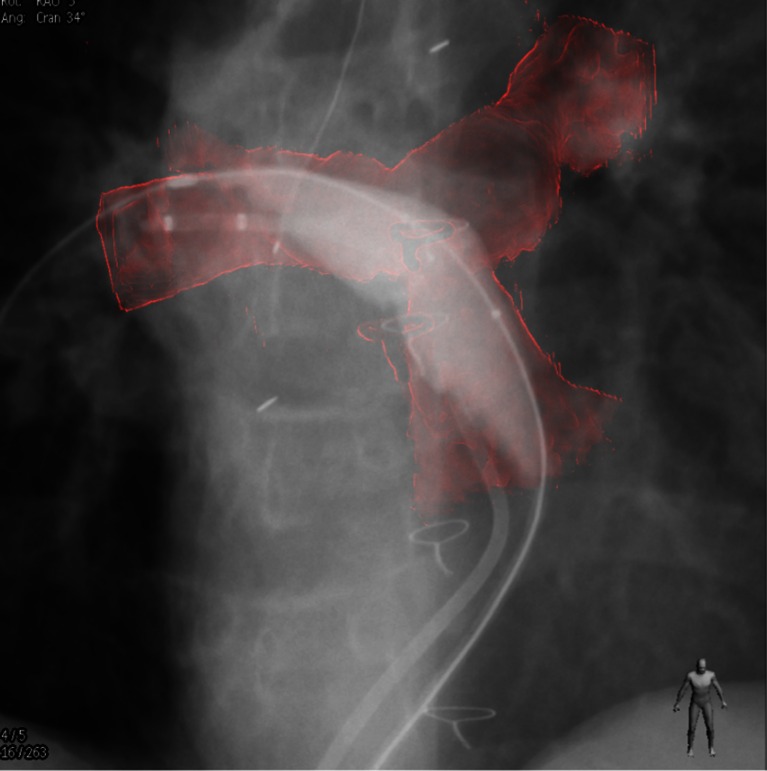
Overlay of 3-dimensional rotational angiogram onto live fluoroscopic images during balloon angioplasty of the right pulmonary artery
References
-
- • Lopez L, Colan SD, Frommelt PC, et al. Recommendations for quantification methods during the performance of a pediatric echocardiogram: a report from the Pediatric Measurements Writing Group of the American Society of Echocardiography Pediatric and Congenital Heart Disease Council. J Am Soc Echocardiogr: Off Publ Am Soc Echocardiogr. May 2010;23(5):465–95; quiz 576-467.This was written by the ASE Pediatric Measurements Writing Group and includes recommended protocols for performing echocardiograms in healthy children and children with congenital heart disease. - PubMed
-
- Jegatheeswaran A, Pizarro C, Caldarone CA, et al. Echocardiographic definition and surgical decision-making in unbalanced atrioventricular septal defect: a Congenital Heart Surgeons' Society multiinstitutional study. Circulation. 2010;122(11 Suppl):S209–15. doi: 10.1161/CIRCULATIONAHA.109.925636. - DOI - PubMed
Publication types
LinkOut - more resources
Full Text Sources