Adhesion of pancreatic cancer cells in a liver-microvasculature mimicking coculture correlates with their propensity to form liver-specific metastasis in vivo
- PMID: 24900957
- PMCID: PMC4037581
- DOI: 10.1155/2014/241571
Adhesion of pancreatic cancer cells in a liver-microvasculature mimicking coculture correlates with their propensity to form liver-specific metastasis in vivo
Abstract
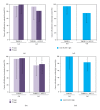
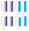
Organ-specific characteristic of endothelial cells (ECs) is crucial for specific adhesion of cancer cells to ECs, which is a key factor in the formation of organ-specific metastasis. In this study, we developed a coculture of TMNK-1 (immortalized liver sinusoidal ECs) with 10T1/2 (resembling hepatic stellate cells) to augment organ-specific characteristic of TMNK-1 and investigated adhesion of two pancreatic cancer cells (MIA-PaCa-2 and BxPC-3) in the culture. MIA-PaCa-2 and BxPC-3 adhesion in TMNK-1+10T1/ 2|coating culture (TMNK-1 monolayer over 10T1/2 layer on collagen coated surface) were similar. However, in TMNK-1+10T1/ 2|gel (coculture on collagen gel surface), MIA-PaCa-2 adhesion was significantly higher than BxPC-3, which was congruent with the reported higher propensity of MIA-PaCa-2 than BxPC-3 to form liver metastasis in vivo. Notably, as compared to BxPC-3, MIA-PaCa-2 adhesion was lower and similar in TMNK-1 only culture on the collagen coated and gel surfaces, respectively. Investigation of the adhesion in the representative human umbilical vein ECs (HUVECs) cultures and upon blocking of surface molecules of ECs revealed that MIA-PaCa-2 adhesion was strongly dependent on the organ-specific upregulated characteristics of TMNK-1 in TMNK-1+10T1/ 2|gel culture. Therefore, the developed coculture would be a potential assay for screening novel drugs to inhibit the liver-microvasculature specific adhesion of cancer cells.
Figures


Similar articles
-
Vascular endothelial growth factor-C secreted by pancreatic cancer cell line promotes lymphatic endothelial cell migration in an in vitro model of tumor lymphangiogenesis.Pancreas. 2007 May;34(4):444-51. doi: 10.1097/mpa.0b13e31803dd307. Pancreas. 2007. PMID: 17446844
-
Cytokines modulate MIA PaCa 2 and CAPAN-1 adhesion to extracellular matrix proteins.Pancreas. 1999 Nov;19(4):362-9. doi: 10.1097/00006676-199911000-00007. Pancreas. 1999. PMID: 10547196
-
Alteration of pancreatic carcinoma and promyeloblastic cell adhesion in liver microvasculature by co-culture of hepatocytes, hepatic stellate cells and endothelial cells in a physiologically-relevant model.Integr Biol (Camb). 2017 Apr 18;9(4):350-361. doi: 10.1039/c6ib00237d. Integr Biol (Camb). 2017. PMID: 28322389
-
Identification of a key molecular regulator of liver metastasis in human pancreatic carcinoma using a novel quantitative model of metastasis in NOD/SCID/gammacnull (NOG) mice.Int J Oncol. 2007 Oct;31(4):741-51. Int J Oncol. 2007. PMID: 17786304
-
Farnesyltransferase inhibitor (L-744,832) restores TGF-beta type II receptor expression and enhances radiation sensitivity in K-ras mutant pancreatic cancer cell line MIA PaCa-2.Oncogene. 2002 Nov 7;21(51):7883-90. doi: 10.1038/sj.onc.1205948. Oncogene. 2002. PMID: 12420225
Cited by
-
Pancreatic Lineage Specifier PDX1 Increases Adhesion and Decreases Motility of Cancer Cells.Cancers (Basel). 2021 Aug 30;13(17):4390. doi: 10.3390/cancers13174390. Cancers (Basel). 2021. PMID: 34503200 Free PMC article.
References
-
- Chaffer CL, Weinberg RA. A perspective on cancer cell metastasis. Science. 2011;331(6024):1559–1564. - PubMed
-
- Irmisch A, Huelsken J. Metastasis: new insights into organ-specific extravasation and metastatic niches. Experimental Cell Research. 2013;319(11):1604–1610. - PubMed
-
- Heikenwalder M, Borsig L. Pathways of metastasizing intestinal cancer cells revealed: how will fighting metastases at the site of cancer cell arrest affect drug development? Future Oncology. 2013;9(1):1–4. - PubMed
-
- Chambers AF, Groom AC, MacDonald IC. Dissemination and growth of cancer cells in metastatic sites. Nature Reviews Cancer. 2002;2(8):563–572. - PubMed
-
- Nguyen DX, Bos PD, Massagué J. Metastasis: from dissemination to organ-specific colonization. Nature Reviews Cancer. 2009;9(4):274–284. - PubMed
Publication types
MeSH terms
LinkOut - more resources
Full Text Sources
Other Literature Sources
Medical

