. 2014:2014:730246.
doi: 10.1155/2014/730246. Epub 2014 May 12.

c-Kit expression, angiogenesis, and grading in canine mast cell tumour: a unique model to study c-Kit driven human malignancies

Affiliations

c-Kit expression, angiogenesis, and grading in canine mast cell tumour: a unique model to study c-Kit driven human malignancies

Rosa Patruno et al. Biomed Res Int. 2014.

Abstract

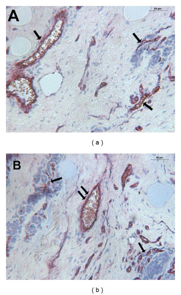
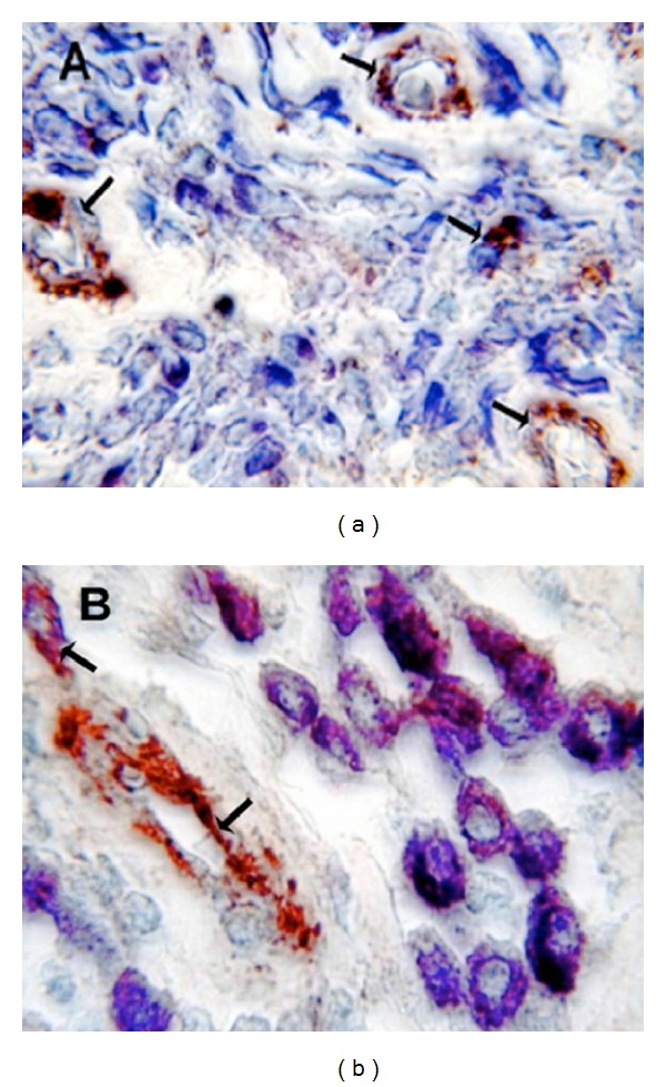
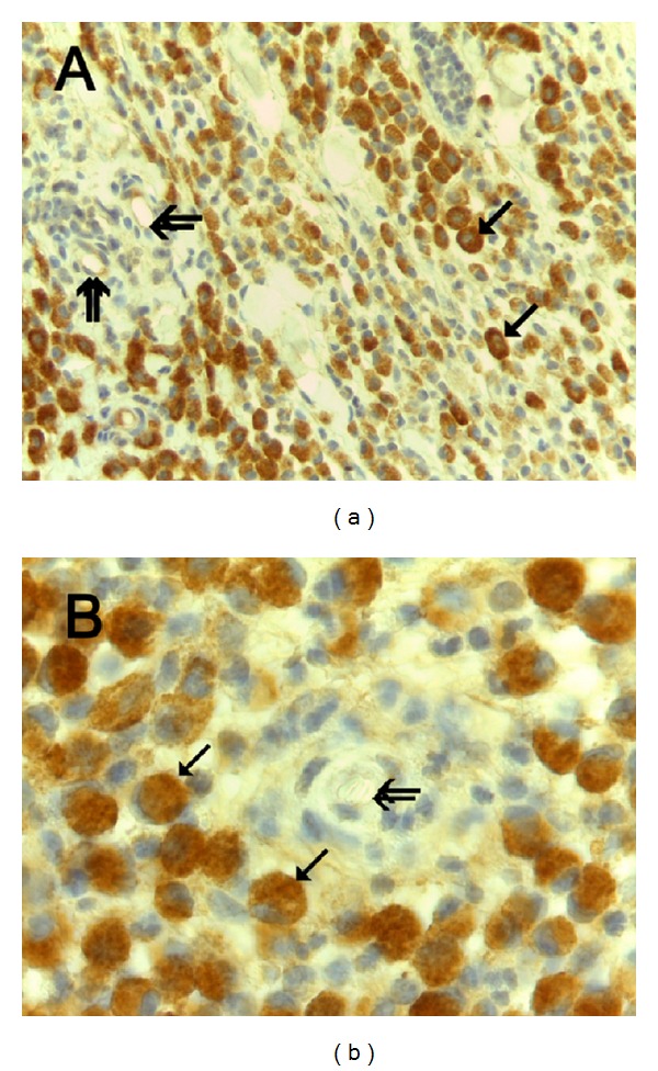
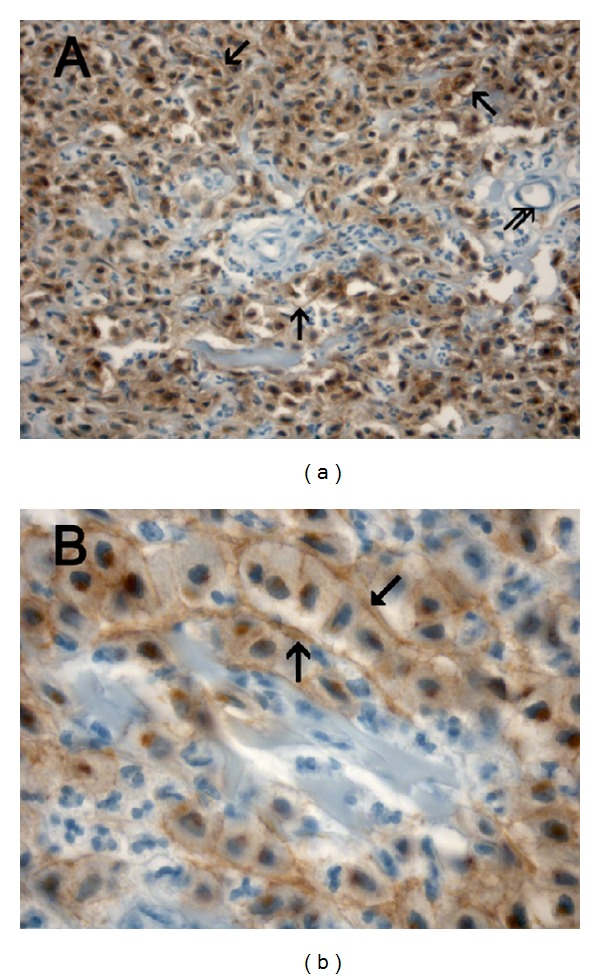
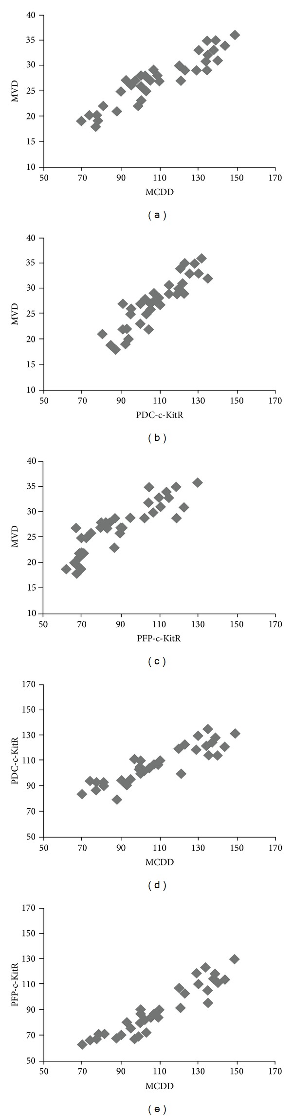
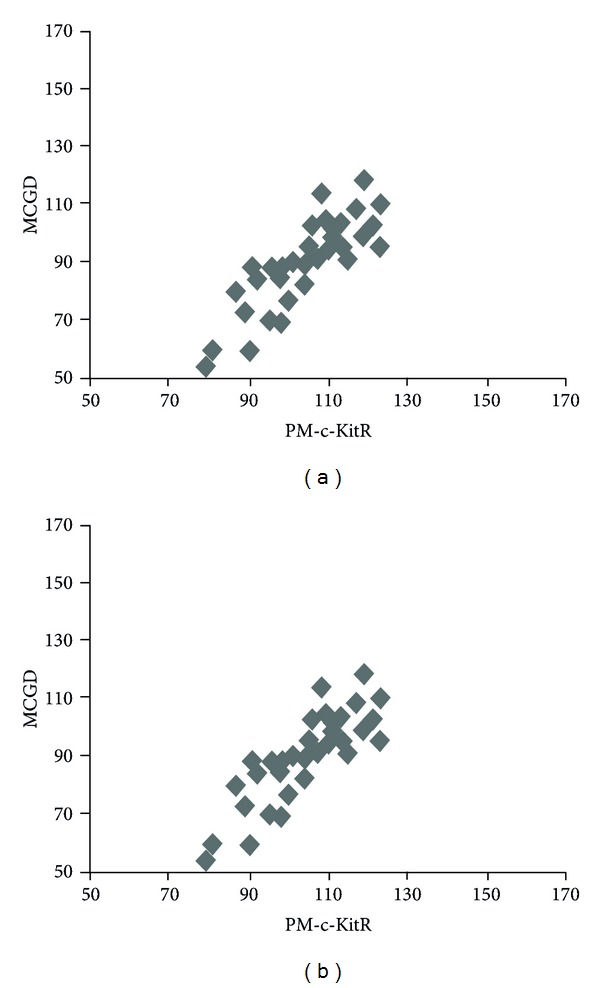
Canine cutaneous mast cell tumour (CMCT) is a c-Kit driven tumour sharing similar c-Kit aberrations found in human gastrointestinal stromal tumour. CMCT is classified into three forms: well- (G1), intermediately (G2) (more benign diseases), and poorly (G3) differentiated (malignant) forms. We assess a correlation between c-Kit status, grading, and angiogenesis in CMCTs to explore their potential significance in humans. C-Kit receptor (c-KitR) expression, microvascular density (MVD), and mast cell granulated and degranulated status density (MCGD and MCDD, resp.) were analyzed in 97 CMCTs, by means of histochemistry, immunohistochemistry double staining, and image analysis system. Data showed that predominantly diffuse cytoplasmic- and predominantly focal paranuclear- (Golgi-like) c-Kit protein (PDC-c-Kit and PFP-c-Kit, resp.) expression correlate with high MVD, G3 histopathological grade, and MCDD. Moreover, predominant cell membrane-c-KitR (PCM-c-KitR) expression status correlates with low MVD, G1-G2 histopathological grade, and MCGD. These findings underline the key role of c-Kit in the biopathology of canine MCTs, indicating a link between aberrant c-Kit expression, increased angiogenesis, and higher histopathological grade. CMCT seems to be a model to study contributions of c-Kit activated MCs in tumour angiogenesis and to evaluate the inhibition of MCs activation by means of c-Kit tyrosine kinase inhibitors, currently translated in humans.


Figures

Figure 1
Figure 1
(a) CMCT with a low MVD. Double staining is performed combining immunohistochemistry with toluidine blue histochemistry. Single arrows indicate blood vessels red-brown immunostained with primary anti-factor VIII-related antigen (FVIII-RA) at ×200 magnification. (b) CMCT with a high MVD. Double staining is performed combining immunohistochemistry toluidine blue histochemistry. Single arrows indicate clusters of blue stained neoplastic mast cells, while double arrows indicate blood vessels red-brown immunostained with a primary anti-FVIII-RA at ×200 magnification.
Figure 2
Figure 2
(a) Poorly differentiated G3 CMCT with high MVD. Double staining is performed combining immunohistochemistry with toluidine blue histochemistry. Many scattered degranulated blue stained mast cells. Single arrows indicate red-brown immunostained microvessels with primary anti-FVIII-RA. Note as an internal positive control the red blood cell in the lumen of microvessel. ×1000 in oil magnification. (b) Well-differentiated G1 CMCT with low MVD. Double staining is performed combining immunohistochemistry with toluidine blue histochemistry. Many scattered granulated red-blue stained mast cells. Single arrows indicate red immunostained microvessels with primary anti-FVIII-RA. Note as an internal positive control the red blood cell in the lumen of microvessel. ×1000 in oil magnification.
Figure 3
Figure 3
(a) Predominantly diffuse cytoplasmic-c-Kit protein (PDC-c-Kit) expression. Immunohistochemistry is performed with primary anti-c-KitR antibody. Single arrows indicate full brown diffuse cytoplasmic immunostained c-KitR. Double arrows indicate microvessels. ×400 magnification. (b) Particular of (a) at even more magnification. Single arrows indicate full brown diffuse cytoplasmic immunostained c-KitR. Double arrows indicate a microvessel. Note as an internal positive control the red blood cells in the lumen of the microvessel. ×1000 in oil magnification.
Figure 4
Figure 4
(a) Predominantly focal paranuclear- (Golgi-like) c-Kit protein (PFP-c-Kit) expression. Immunohistochemistry is performed with primary anti-c-KitR antibody. Single arrows indicate focal brown paranuclear cytoplasmic immunostained c-KitR. Double arrows indicate a vessel. Note as an internal positive control the red blood cells in the lumen of the vessel. ×400 magnification. (b) Particular of (a) at even more magnification. Immunohistochemistry is performed with primary anti-c-KitR antibody. Single arrows indicate brown paranuclear cytoplasmic (Golgi-like) immunostained c-KitR. Double arrows indicate microvessels. Note as an internal positive control the red blood cells in the lumen of microvessels. ×1000 in oil magnification.
Figure 5
Figure 5
(a) Predominant cell membrane-c-KitR (PCM-c-KitR) expression. Immunohistochemistry is performed with primary anti-c-KitR antibody. Single arrows indicate threadlike cell membrane brown immunostained c-KitR. Double arrows indicate a microvessel. Note as an internal positive control a red blood cell in the lumen of the microvessel. ×400 magnification. (b) Particular of (a) at even more magnification. Immunohistochemistry is performed with primary anti-c-KitR antibody. Single arrows indicate cell membrane brown immunostained c-KitR. ×1000 in oil magnification.
Figure 6
Figure 6
Panel shows the correlations between MVD, MCDD, PFP-c-KitR, and PDC-c-KitR by Pearson's (r) analysis in G3 CMCTs subgroup.
Figure 7
Figure 7
Panel shows the correlations between MCGD and PM-c-KitR by Pearson's (r) analysis in G1 and G2 CMCTs subgroups.

References

    1. Kitamura Y, Hirotab S. Kit as a human oncogenic tyrosine kinase. Cellular and Molecular Life Sciences. 2004;61(23):2924–2931. - PMC - PubMed
    1. London CA, Galli SJ, Yuuki T, Hu Z, Helfand SC, Geissler EN. Spontaneous canine mast cell tumors express tandem duplications in the proto-oncogene c-kit. Experimental Hematology. 1999;27(4):689–697. - PubMed
    1. London CA, Kisseberth WC, Galli SJ, Geissler EN, Helfand SC. Expression of stem cell factor receptor (c-kit) by the malignant mast cells from spontaneous canine mast cell tumours. Journal of Comparative Pathology. 1996;115(4):339–414. - PubMed
    1. Takeuchi Y, Fujino Y, Watanabe M, et al. Validation of the prognostic value of histopathological grading or c-kit mutation in canine cutaneous mast cell tumours: a retrospective cohort study. Veterinary Journal. 2013;196(3):492–498. - PubMed
    1. Kim KH, Nelson SD, Kim DH, et al. Diagnostic relevance of overexpressions of PKC-θ and DOG-1 and KIT/PDGFRA gene mutations in extragastrointestinal stromal tumors: a Korean six-centers study of 28 cases. Anticancer Research. 2012;32(3):923–937. - PubMed

Publication types

MeSH terms

Substances

LinkOut - more resources