. 2014 Dec;20(23-24):3228-40.
doi: 10.1089/ten.TEA.2013.0729.

Improved peripheral nerve regeneration using acellular nerve allografts loaded with platelet-rich plasma

Affiliations

Improved peripheral nerve regeneration using acellular nerve allografts loaded with platelet-rich plasma

Canbin Zheng et al. Tissue Eng Part A. 2014 Dec.

Abstract

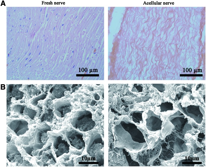
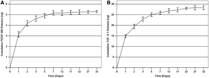
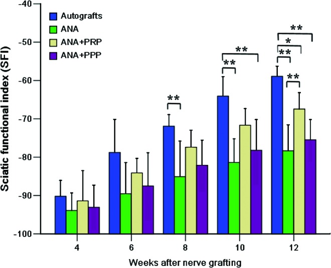
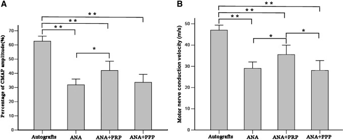
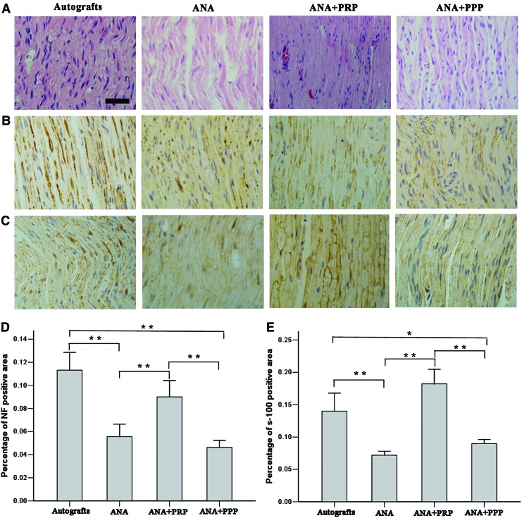
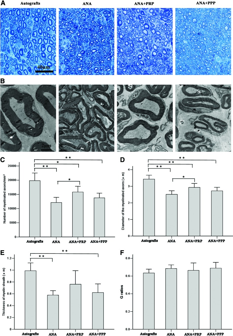
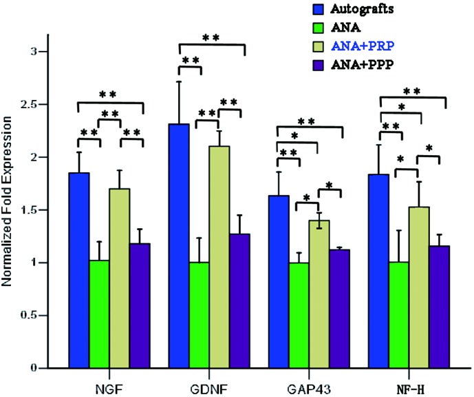
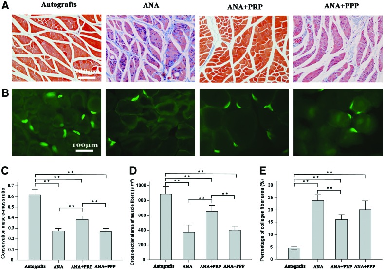
Acellular nerve allografts (ANAs) behave in a similar manner to autografts in supporting axonal regeneration in the repair of short peripheral nerve defects but fail in larger defects. The objective of this article is to evaluate the effect of ANA supplemented with platelet-rich plasma (PRP) to improve nerve regeneration after surgical repair and to discuss the mechanisms that underlie this approach. Autologous PRP was obtained from rats by double-step centrifugation and was characterized by determining platelet numbers and the release of growth factors. Forty-eight Sprague-Dawley rats were randomly divided into 4 groups (12/group), identified as autograft, ANA, ANA loaded with PRP (ANA+PRP), and ANA loaded with platelet-poor plasma (PPP, ANA+PPP). All grafts were implanted to bridge long-gap (15 mm) sciatic nerve defects. We found that PRP with a high platelet concentration exhibited a sustained release of growth factors. Twelve weeks after surgery, the autograft group displayed the highest level of reinnervation, followed by the ANA+PRP group. The ANA+PRP group showed a better electrophysiology response for amplitude and conduction velocity than the ANA and ANA+PPP groups. Based on histological evaluation, the ANA+PRP and autograft groups had higher numbers of regenerating nerve fibers. Quantitative real-time polymerase chain reaction (qRT-PCR) demonstrated that PRP boosted expression of neurotrophins in the regenerated nerves. Moreover, the ANA+PRP and autograft groups showed excellent physiological outcomes in terms of the prevention of muscle atrophy. In conclusion, ANAs loaded with PRP as tissue-engineered scaffolds can enhance nerve regeneration and functional recovery after the repair of large nerve gaps nearly as well as autografts.


Figures

<b>FIG. 1.</b>
FIG. 1.
Morphological observations of the acellular nerves. (A) A longitudinal section of the fresh nerve and acellular nerve by hematoxylin and eosin (H&E) staining. (B) Scanning electron microscopy images of the fresh nerve and acellular nerve. Compared with the fresh nerve, the components within cells, the myelin and the axon have been removed, whereas the endoneurial basal laminae remain intact. Color images available online at www.liebertpub.com/tea
<b>FIG. 2.</b>
FIG. 2.
The cumulative in vitro release of (A) platelet-derived growth factor (PDGF)-BB and (B) transforming growth factor-β1 (TGF-β1) from platelet-rich plasma (PRP) gels after leaching in phosphate-buffered saline (mean±standard deviation [SD]; n=3 for one batch). Both gels show a burst release of approximately 24 h, with a subsequent gradual release rate for 7 days.
<b>FIG. 3.</b>
FIG. 3.
A plot showing the sciatic functional index (SFI) evolution of the four grafted groups at 4, 6, 8, 10, and 12 weeks after nerve grafting. n=8 for each group. *p<0.05, **p<0.01. Color images available online at www.liebertpub.com/tea
<b>FIG. 4.</b>
FIG. 4.
Representative compound muscle action potential (CMAP) recordings for the four groups at 12 weeks after nerve grafting. (A) Histograms showing the CMAP recovery ratio and (B) motor nerve conduction velocity detected at the injured side. Error bars correspond to mean±SD (n=8 for each group). *p<0.05, **p<0.01.
<b>FIG. 5.</b>
FIG. 5.
Longitudinal sections of the regenerated nerve tissues and histological analysis for the four groups at week 12 after nerve grafting. (A) The longitudinal sections of the segments distal to the nerve grafts analyzed by H&E staining. (B) Immunostaining with anti-neurofilament antibody in longitudinal sections. (C) Immunostaining for the Schwann cell marker S-100 in the longitudinal sections. (D) Statistical analysis of the neurofilament-positive area for each group. (E) Statistical analysis for the S-100-positive area for each group. There were significant increases in the numbers of positively stained neurofilaments and S-100 for the autografts and acellular nerve allograft (ANA)+PRP groups but not for the ANA and ANA+platelet-poor plasma (PPP) groups. Error bars correspond to mean±SD (n=8 for each group). *p<0.05, **p<0.01. Scale bar=50 μm. Color images available online at www.liebertpub.com/tea
<b>FIG. 6.</b>
FIG. 6.
Transverse sections of the midpoint of the harvested grafts from the four groups at week 12 after nerve grafting. (A) Staining with toluidine blue shows the regenerated axons. The autografts and the ANA+PRP group appear to have more regenerating nerve fibers than the other two groups. (B) Transmission electron micrographs demonstrate an increased amount of axon bundling and myelination in the autograft and ANA+PRP groups compared with the ANA and ANA+PPP groups. Comparisons of (C) the density, (D) axonal diameter, (E) thickness of the myelin sheaths, and (F) G ratios of the myelinated nerve fibers. Error bars correspond to mean±SD (n=8 for each group). *p<0.05, **p<0.01. Color images available online at www.liebertpub.com/tea
<b>FIG. 7.</b>
FIG. 7.
The mRNA expression for nerve growth factor (NGF), glial-derived growth factor (GDNF), growth-associated protein 43 (GAP43), and neurofilament heavy (NF-H), as measured by quantitative real-time polymerase chain reaction (qRT-PCR), in the regenerating nerves taken from the four groups at 12 weeks after nerve grafting. The mRNA expression was calculated by the 2−ΔΔCT method using GDPHA as the reference gene. The relative mRNA levels were represented as the ratios by comparing the expression of each group with that of the ANA group (designated as 1). n=4 for each group, *p<0.05, **p<0.01. Color images available online at www.liebertpub.com/tea
<b>FIG. 8.</b>
FIG. 8.
Light micrographs of (A) Masson trichrome staining of transverse sections of the triceps surae muscles and (B) cholinesterase staining of the motor endplate on longitudinal sections of the triceps surae muscles from the sides of the rats that underwent operation, recorded at 12 weeks after nerve grafting. Histograms showing (C) conservation muscle–mass ratio, (D) cross-sectional area of the muscle fibers, and (E) average percentage of collagen fiber area for the triceps surae muscle samples from the sides that underwent the operation. Error bars correspond to mean±SD (n=8 for each group). *p<0.05, **p<0.01. Color images available online at www.liebertpub.com/tea

References

    1. Huang W., Begum R., Barber T., Ibba V., Tee N.C., Hussain M., Arastoo M., Yang Q., Robson L.G., Lesage S., Gheysens T., Skaer N.J., Knight D.P., and Priestley J.V.Regenerative potential of silk conduits in repair of peripheral nerve injury in adult rats. Biomaterials 33,59, 2012 - PubMed
    1. Shen C.C., Yang Y.C., and Liu B.S.Peripheral nerve repair of transplanted undifferentiated adipose tissue-derived stem cells in a biodegradable reinforced nerve conduit. J Biomed Mater Res A 100,48, 2012 - PubMed
    1. Kim B.S., Yoo J.J., and Atala A.Peripheral nerve regeneration using acellular nerve grafts. J Biomed Mater Res A 68,201, 2004 - PubMed
    1. Yang L.M., Liu X.L., Zhu Q.T., Zhang Y., Xi T.F., Hu J., He C.F., and Jiang L.Human peripheral nerve-derived scaffold for tissue-engineered nerve grafts: histology and biocompatibility analysis. J Biomed Mater Res B Appl Biomater 96,25, 2011 - PubMed
    1. Giusti G., Willems W.F., Kremer T., Friedrich P.F., Bishop A.T., and Shin A.Y.Return of motor function after segmental nerve loss in a rat model: comparison of autogenous nerve graft, collagen conduit, and processed allograft (AxoGen). J Bone Joint Surg Am 94,410, 2012 - PubMed

Publication types

LinkOut - more resources