. 2015 Feb;5(1):35-44.
doi: 10.1089/brain.2014.0251. Epub 2014 Jul 31.

Evolution of functional connectivity of brain networks and their dynamic interaction in temporal lobe epilepsy

Affiliations

Evolution of functional connectivity of brain networks and their dynamic interaction in temporal lobe epilepsy

Victoria L Morgan et al. Brain Connect. 2015 Feb.

Abstract

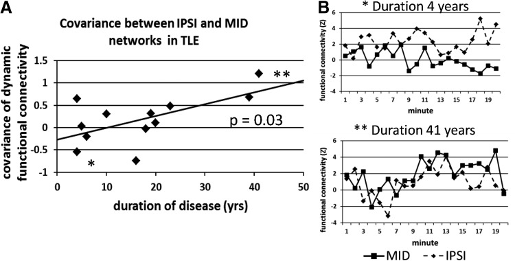
This study presents a cross-sectional investigation of functional networks in temporal lobe epilepsy (TLE) as they evolve over years of disease. Networks of interest were identified based on a priori hypotheses: the network of seizure propagation ipsilateral to the seizure focus, the same regions contralateral to seizure focus, the cross hemisphere network of the same regions, and a cingulate midline network. Resting functional magnetic resonance imaging data were acquired for 20 min in 12 unilateral TLE patients, and 12 age- and gender-matched healthy controls. Functional changes within and between the four networks as they evolve over years of disease were quantified by standard measures of static functional connectivity and novel measures of dynamic functional connectivity. The results suggest an initial disruption of cross-hemispheric networks and an increase in static functional connectivity in the ipsilateral temporal network accompanying the onset of TLE seizures. As seizures progress over years, the static functional connectivity across the ipsilateral network diminishes, while dynamic functional connectivity measures show the functional independence of this ipsilateral network from the network of midline regions of the cingulate declines. This implies a gradual breakdown of the seizure onset and early propagation network involving the ipsilateral hippocampus and temporal lobe as it becomes more synchronous with the network of regions responsible for secondary generalization of the seizures, a process that may facilitate the spread of seizures across the brain. Ultimately, the significance of this evolution may be realized in relating it to symptoms and treatment outcomes of TLE.

Keywords: brain; functional connectivity; functional magnetic resonance imaging; network; seizure propagation; temporal lobe epilepsy.


Figures

<b>FIG. 1.</b>
FIG. 1.
Network diagram. The eight regions of interest are shown as circles, and each label is provided. The four networks identified from these regions in one individual subject are each shown in a different color: IPSI in blue and the regions on the coronal brain slice are also shown in blue, CONTRA in red with regions on the coronal brain slice shown in red, CROSS in purple, and MID in green with two of the four nodes (cingulate and precuneus) shown in green on the axial brain slice. See “Regions of interest” section for more detailed descriptions of regions and networks. HIPI, HIPC: hippocampus ipsilateral, contralateral; INSI, INSC: insula ipsilateral, contralateral; THALI, THALC: thalamus ipsilateral, contralateral; CING: mid cingulate gyrus; PREC: precuneus; IPSI: ipsilateral network; CONTRA: contralateral network; CROSS: cross hemispheric network; MID: midline cingulate network.
<b>FIG. 2.</b>
FIG. 2.
Network functional connectivity processing steps beginning with the eight region by eight region correlation matrix derived from each of the two 10 min fMRI imaging series (A). Dynamic and between network functional connectivity processing steps beginning with the eight region by eight region correlation matrix derived from each minute of the 20 min of fMRI acquisition (B). These steps represent processing as performed on each individual subject. fMRI, functional magnetic resonance imaging.
<b>FIG. 3.</b>
FIG. 3.
Functional connectivity within the CROSS network versus duration of disease in years. The TLE subjects are shown in closed diamonds and the controls are indicated in open circles. The healthy controls are plotted at the same duration of disease as their corresponding age- and gender-matched TLE patient. The trendline represents the linear fit of the TLE data of patients with duration of disease greater than 5 years. TLE, temporal lobe epilepsy.
<b>FIG. 4.</b>
FIG. 4.
The functional connectivity within the IPSI network versus duration of disease in TLE (A). Linear trendline is included. The functional connectivity over the 20 min fMRI acquisition is shown in (B) for two different pairs of subjects: TLE with 4 years' duration and matched control (top, point in A indicated by *) and TLE with 41 years' duration and matched control (bottom, point in A indicated by **).
<b>FIG. 5.</b>
FIG. 5.
The variability of the functional connectivity of the MID network over the 20 min fMRI acquisition versus duration of disease in TLE. Linear trendline is included.
<b>FIG. 6.</b>
FIG. 6.
The covariance of the functional connectivity of the IPSI and MID networks versus duration of disease in TLE (A). Linear trendline is included. The functional connectivity over the 20 min fMRI acquisition in each network is shown in (B) for two different subjects: TLE with 4 years' duration (top, point in A indicated by *), and TLE with 41 years' duration (bottom, point in A indicated by **).

References

    1. Allen EA, Damaraju E, Plis SM, Erhardt EB, Eichele T, Calhoun VD. 2014. Tracking whole-brain connectivity dynamics in the resting state. Cereb Cortex 24:663–676 - PMC - PubMed
    1. Bernasconi N, Duchesne S, Janke A, Lerch J, Collins DL, Bernasconi A. 2004. Whole-brain voxel-based statistical analysis of gray alter and white matter in temporal lobe epilepsy. Neuroimage 23:717–723 - PubMed
    1. Bettus G, Bartolomei F, Confort-Gouny S, Guedj E, Chauvel P, Cozzone PJ, et al. . 2010. Role of resting state functional connectivity MRI in presurgical investigation of mesial temporal lobe epilepsy. J Neurol Neurosurg Psychiatry 81:1147–1154 - PubMed
    1. Bettus G, Guedj E, Joyeux F, Confort-Gouny S, Soulier E, Laguitton V, et al. . 2009. Decreased basal fMRI functional connectivity in epileptogenic networks and contralateral compensatory mechanisms. Hum Brain Mapp 30:1580–1591 - PMC - PubMed
    1. Blauwblomme T, David O, Minotti L, Job AS, Chassagnon S, Hoffman D, et al. . 2013. Prognostic value of insular lobe involvement in temporal lobe epilepsy: a stereoelectroencephalographic study. Epilepsia 54:1658–1667 - PubMed

Publication types