. 2014 Jun 5;5(6):e1274.
doi: 10.1038/cddis.2014.232.

Retinoblastoma protein (RB) interacts with E2F3 to control terminal differentiation of Sertoli cells

Affiliations

Retinoblastoma protein (RB) interacts with E2F3 to control terminal differentiation of Sertoli cells

E Rotgers et al. Cell Death Dis. .

Abstract

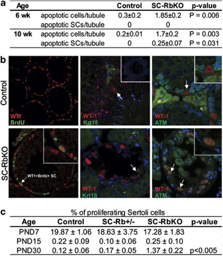
The retinoblastoma protein (RB) is essential for normal cell cycle control. RB function depends, at least in part, on interactions with the E2F family of DNA-binding transcription factors (E2Fs). To study the role of RB in the adult testis, a Sertoli cell (SC)-specific Rb knockout mouse line (SC-RbKO) was generated using the Cre/loxP recombination system. SC-RbKO mice exhibited an age-dependent testicular atrophy, impaired fertility, severe SC dysfunction, and spermatogenic defects. Removal of Rb in SC induced aberrant SC cycling, dedifferentiation, and apoptosis. Here we show that E2F3 is the only E2F expressed in mouse SCs and that RB interacts with E2F3 during mouse testicular development. In the absence of RB, the other retinoblastoma family members p107 and p130 began interacting with E2F3 in the adult testes. In vivo silencing of E2F3 partially restored the SC maturation and survival as well as spermatogenesis in the SC-RbKO mice. These results point to RB as a key regulator of SC function in adult mice and that the RB/E2F3 pathway directs SC maturation, cell cycle quiescence, and RB protects SC from apoptosis.


Figures

Figure 1
Figure 1
RB loss in SC leads to spermatogenic failure in adult mice. (a) Reproductive tracts of 10-week-old control and SC-specific Rb knockout (SC-RbKO) male mice showing gross atrophy of SC-RbKO testes. (b) A significant reduction in relative testis weights of SC-RbKO 10–12-week-old mice in comparison with control and SC-Rb+/− testes. (c) Testis sections from control and SC-RbKO mice (10-week old) stained with PAS. Round spermatids in symplasts, dashed black circles; SCs, black arrows. Scale bars, 50 and 25 μm. (d) Cauda epididymal sperm counts from 10–12-week-old SC-RbKO versus control mice showing a significant decrease of sperm counts. In addition, 45% of the sperm are headless in SC-RbKO. Error bars, S.E.M.; P<0.05, P<0.001. (e) H&E staining showing control cauda epididymis full of spermatozoa (10-week old). In the SC-RbKO epididymis, there are numerous exfoliated immature germ cells and the mature sperm are less abundant. Scale bar, 25 μm
Figure 2
Figure 2
Loss of RB results in apoptosis of germ cell and SCs and re-entry of SCs to mitotic cell cycle. (a) Quantification of apoptotic germ cells (intratubular cells positive for TUNEL only) and SCs (intratubular cells positive for both WT1 and TUNEL) in control and SC-RbKO testes at the ages of 6 and 10 weeks. (n=4/group). Increased germ cell apoptosis was detected already at the age of 6 weeks in the SC-RbKO testes, and by the age of 10 weeks also the SC begin to undergo apoptosis in the SC-RbKO. (b) Left panels: Analysis of BrdU incorporation was performed in control (upper) and SC-RbKO (lower) testes at PND30. Double immunofluorescence labelling of WT-1 (red) and BrdU (green) revealed that a significant number of adult RB-deficient SCs maintain DNA synthesis (inset). Middle panels: Double immunofluorescent labelling of cytokeratin 18 (Krt18, green) and WT-1 (red) in testes from control and SC-RbKO mice (12-week old). Positive staining of Krt18, a marker of immature SCs, in RbKO-SCs (WT-1+). Inset: Krt18 in PND1 mouse testis, positive control. Permeabilization in TE buffer produces unspecific signal in the sex body of the round spermatids. Right panels: Double immunofluorescence labelling of phosphorylated-ATM (pATM, green) and WT-1 (red) in testes from control and SC-RbKO mice (12-week old). In the wild-type SC nuclei, phospho-ATM is in the perinuclear area, whereas in the SC-RbKO SC pATM signal is localized to the nucleus. Inset: close-up on pATM+ SC. Magnifications are × 200 and × 00. (c) Quantification of proliferating SCs (WT1+/BrdU+) in mice testes shows comparable proliferative rate of WT and SC-RbKO SC during the normal proliferative time window but a cell-cycle re-entry at the PND30 in the SC-RbKO SCs (n=4/group)
Figure 3
Figure 3
E2F3 in mouse SCs is developmentally controlled by the RB family. (a) Immunolocalization of E2F3 in SC (white arrows), preleptotene spermatocytes (black arrowheads), and spermatogonia (Spg, white arrowheads) of adult control and SC-RbKO mice. NC: negative control, secondary antibody only. Scale bar, 25 μm. (b) mRNA levels of E2f3 in control and SC-RbKO mice testes at 6 and 10–12 weeks of age relative to housekeeping genes (L19 and Ppia) and normalized to the control group. (c) Both E2F3 isoforms a and b are expressed at high levels in 10–12-week-old control (WT) and KO testes. E2F3++TM4; E2F3-overexpressing TM4 mouse-SC line (positive control); liver, negative control. (d) Co-immunoprecipitation using E2F3 antibody from whole-testis lysate from PND10 and PND40 control and SC-RbKO mice. WB analysis of RB family members shows that E2F3 interacts with RB at PND10 in the wild-type testes. Following RB deletion, E2F3 forms complexes with both p107 and p130 in the PND40 SC-RbKO testes. NC: negative control is immunoprecipitation with the same E2F3 antibody in the presence of a specific blocking peptide (sc-878 P)
Figure 4
Figure 4
Expression patterns of RB family members p107 and p130 in the WT and SC-RbKO testes. (a) p107 was detected only in pachytene spermatocytes in 12-week-old control testes; however, in SC-RbKO mouse testes positive p107 signal was also detected in a proportion of SC. SCs, black arrowheads; pachytene spermatocytes, black circles. SCs with p107 signal, asterisk. (b) p130 was detected in SC, in both control and SC-RbKO mouse testes. SCs, black arrowheads. (c and d) p130 and p107 mRNA levels in SC-RbKO testes versus control at the ages of 6 and 10–12 weeks. n=4/group
Figure 5
Figure 5
Partial rescue of spermatogenesis in SC-RbKO mouse testes following in vivo E2F3 silencing. (a) A significant recovery of testis weight following in vivo knockdown of E2F3ab in the 12-week-old SC-RbKO testes. Control: non-transfected WT testis, SC-RbKO: non-transfected mutant, pSuperEGFP: mutant transfected with empty pSUPER.gfp/neo plasmid, SiE2F3ab: mutant transfected with shRNA-E2F3ab plasmid. Values are mean±S.E.M. of n=4. (b) GFP expression introduced by the in vivo transfection was confirmed 10 weeks after injection by a squash preparation of seminiferous tubule segment under fluorescence microscopy containing transfected SCs (red arrowhead). (c) H&E staining revealed normal spermatogenesis in control mouse testes at the age of 12 weeks (upper left panel) and disorganized seminiferous tubules in testes from SC-RbKO (upper middle panel) and SC-RbKO-transfected with control plasmid (KO/pSuperEGFP) at the same age (upper right panel). Testicular sections of SC-RbKO mice transfected with shRNA-E2F3ab plasmid (lower panels) show elongated spermatids and a restored seminiferous tubule architecture. (d) Testis architecture, ectoplasmic specializations (espin: green, left panels) and tight junctions (claudin11: red, right panels) were restored in SC-RbKO after knockdown of E2F3 (Magnification × 400)
Figure 6
Figure 6
Hypothetical model of the RB family and E2F3 dynamics during mouse SC development. Wild-type: In the juvenile mouse SC, which are actively proliferating and have not yet formed an intact BTB, RB and E2F3 interact physically, to either activate or repress E2F3-mediated gene expression. In the terminally differentiated SCs, there is a tightly regulated balance of RB and E2F3 action. The RB family member p130 is expressed in wild-type SC in addition to RB. SC-RbKO: The juvenile SC proliferate normally and exhibit no apparent phenotype beyond altered gene expression profile in the absence of RB. Neither RB nor p130 interact with E2F3 in the juvenile SC-RbKO SC. The adult SC-RbKO SC gradually resume proliferation, become dedifferentiated, apoptotic, and fail to support spermatogenesis. Simultaneously, SC-RbKO SC assume an ectopic expression of the RB family member p107, and p107 and p130 both begin to interact with E2F3; possibly attempting to suppress a deregulated E2F3 action. Rescued SC-RbKO: E2F3 was knocked down in vivo using a shRNA in PND15 SC-RbKO testes to assess the contribution of E2F3 to the SC-RbKO phenotype. This resulted in partial rescue of the SC-RbKO phenotype with a patchy restoration of spermatogenesis and seminiferous tubule architecture

Similar articles

Cited by

References

    1. 1Classon M, Harlow E. The retinoblastoma tumour suppressor in development and cancer. Nat Rev Cancer 2002; 2: 910–917. - PubMed
    1. 2Burkhart DL, Sage J. Cellular mechanisms of tumour suppression by the retinoblastoma gene. Nat Rev Cancer 2008; 8: 671–682. - PMC - PubMed
    1. 3Yan W, Kero J, Suominen J, Toppari J. Differential expression and regulation of the retinoblastoma family of proteins during testicular development and spermatogenesis: roles in the control of germ cell proliferation, differentiation and apoptosis. Oncogene 2001; 20: 1343–1356. - PubMed
    1. 4Yan W, West A, Toppari J, Lahdetie J. Stage-specific expression and phosphorylation of retinoblastoma protein (pRb) in the rat seminiferous epithelium. Mol Cell Endocrinol 1997; 132: 137–148. - PubMed
    1. 5Clarke AR, Maandag ER, van Roon M, van der Lugt NM, van der Valk M, Hooper ML et al. Requirement for a functional Rb-1 gene in murine development. Nature 1992; 359: 328–330. - PubMed

Publication types