. 2014 Jun 5;10(6):e1004149.
doi: 10.1371/journal.ppat.1004149. eCollection 2014 Jun.

Dusp3 and Psme3 are associated with murine susceptibility to Staphylococcus aureus infection and human sepsis

Affiliations

Dusp3 and Psme3 are associated with murine susceptibility to Staphylococcus aureus infection and human sepsis

Qin Yan et al. PLoS Pathog. .

Erratum in

  • PLoS Pathog. 2014 Jun;10(6):e1004259. Thaden, Joshua [corrected to Thaden, Joshua T]

Abstract

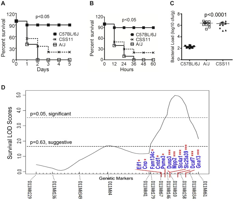
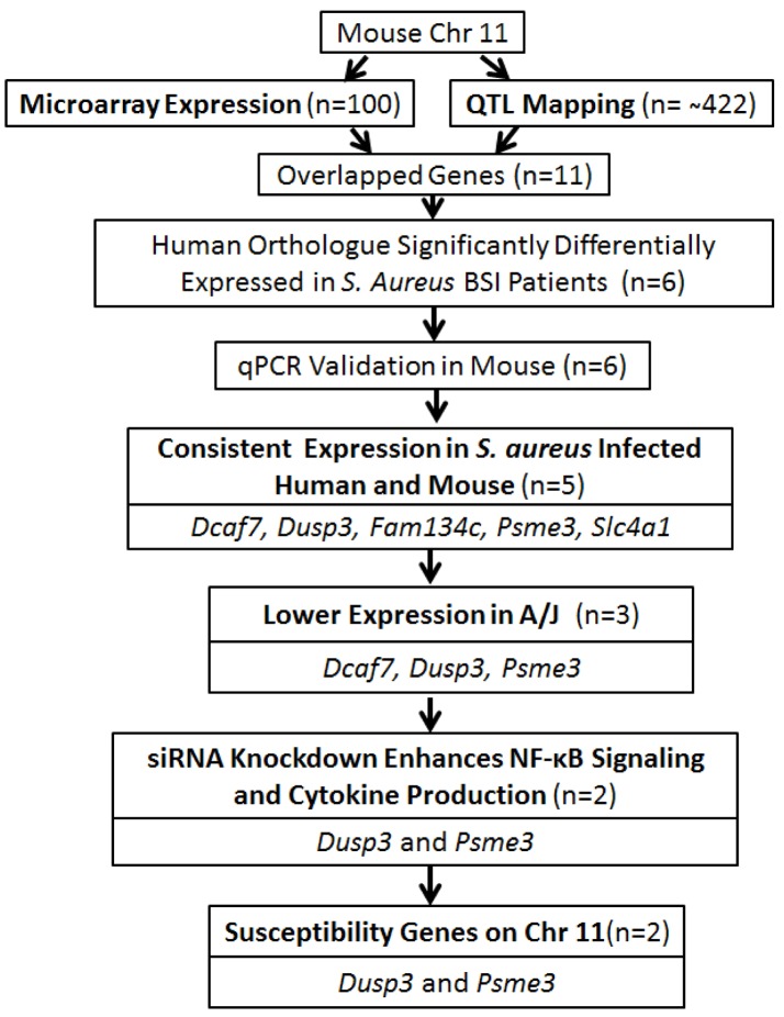
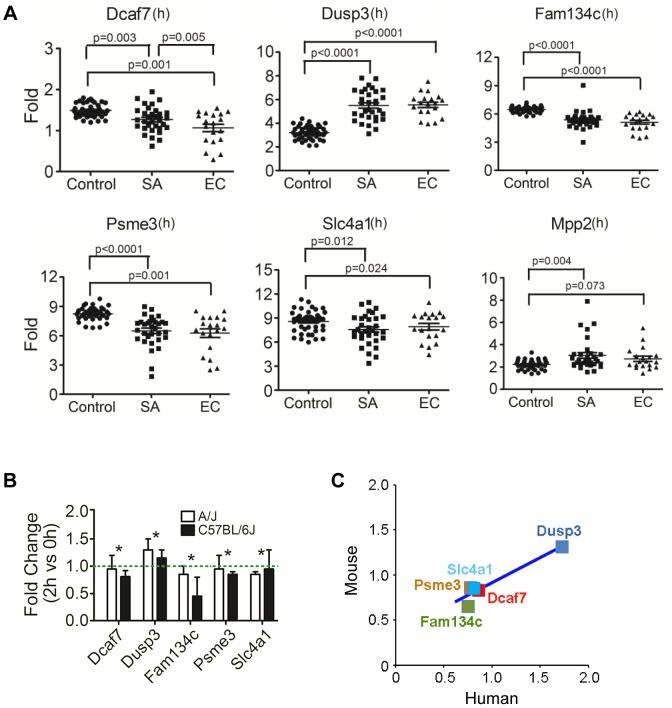
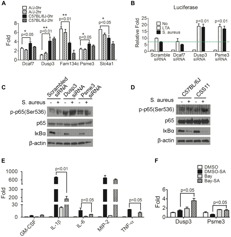
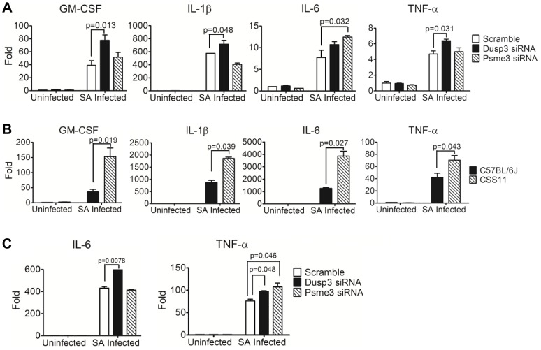
Using A/J mice, which are susceptible to Staphylococcus aureus, we sought to identify genetic determinants of susceptibility to S. aureus, and evaluate their function with regard to S. aureus infection. One QTL region on chromosome 11 containing 422 genes was found to be significantly associated with susceptibility to S. aureus infection. Of these 422 genes, whole genome transcription profiling identified five genes (Dcaf7, Dusp3, Fam134c, Psme3, and Slc4a1) that were significantly differentially expressed in a) S. aureus -infected susceptible (A/J) vs. resistant (C57BL/6J) mice and b) humans with S. aureus blood stream infection vs. healthy subjects. Three of these genes (Dcaf7, Dusp3, and Psme3) were down-regulated in susceptible vs. resistant mice at both pre- and post-infection time points by qPCR. siRNA-mediated knockdown of Dusp3 and Psme3 induced significant increases of cytokine production in S. aureus-challenged RAW264.7 macrophages and bone marrow derived macrophages (BMDMs) through enhancing NF-κB signaling activity. Similar increases in cytokine production and NF-κB activity were also seen in BMDMs from CSS11 (C57BL/6J background with chromosome 11 from A/J), but not C57BL/6J. These findings suggest that Dusp3 and Psme3 contribute to S. aureus infection susceptibility in A/J mice and play a role in human S. aureus infection.


Conflict of interest statement

VGF served as Chair of V710 Scientific Advisory Committee (Merck), has received grant support from Cerexa, Pfizer, Advanced Liquid Logic, MedImmune, has been a paid consultant for Merck, Astellas, Affinium, Bayer, Theravance, Cubist, Cerexa, Durata, Pfizer, NovaDigm, Novartis, Medicines Company, Biosynexus, MedImmune, and Inimex, and has received honoraria from Merck, Astellas, Cubist, Pfizer, Theravance, and Novartis. JL is employed by Quintiles Innovations. This does not alter our adherence to all PLOS policies on sharing data and materials.

Figures

Figure 1
Figure 1. Chromosome substitution strain 11 was susceptible to S. aureus infection, and QTL mapping found eleven putative candidate genes on Chr11.
(A) CSS11 was susceptible to S. aureus peritoneal sepsis. C57BL/6J, A/J, or CSS11 mice were injected (i.p.) with S. aureus Sanger 476 at 107 CFU/g (n = 10 for each strain). Comparison of survival curves was performed by Mann-Whitney u test. The difference between C57BL/6J and CSS11 mice was significant (p<0.05). (B) CSS11 was susceptible to S. aureus intravenous sepsis. C57BL/6J, A/J, or CSS11 were intravenous injected with S. aureus Sanger 476 at 2×10⁶ CFU/g (n = 10 for each strain). Comparison of survival curves was performed by Mann-Whitney u test. The difference between C57BL/6J and CSS11 mice was significant (p<0.05). (C) Bacterial load in kidneys were significantly higher in CSS11 mice after S. aureus injection. C57BL/6J, A/J and CSS11 were injected (i.p.) with S. aureus Sanger 476 at 107 CFU/g and euthanized 24 hours post infection (n = 10 for each group). The bacterial load in CSS11 kidneys were significantly higher than C57BL/6J (2.0±1.32×10⁶ CFU/ml vs 200±158 CFU/ml, p<0.0001). (D) Chromosome 11 LOD score plot for susceptibility to S. aureus in F2 intercross mice (F1 [C11A]×F1 [C11A]). Six to eight-week-old intercross mice were injected i.p. with 107 CFU/g S. aureus Sanger 476 and observed every 8 hours continuously for 5 days. Thresholds for significant (p = 0.05) and suggestive (p = 0.63) linkage are indicated by the horizontal dashed lines. LOD score was determined by the J/qtl permutation test using 1,000 permuted data sets. The microsatellite markers for determining genotypes of F2 intercross mice are marked along the X-axis. The differentially expressed genes are indicated. Genes identified within significant or suggestive QTL were indicated with *** or *, respectively. Footnote: Figure 1A and 1C have been presented in Ahn S, et al., 2010 PLoS Pathogens, e1001088.
Figure 2
Figure 2. Overall strategy for identifying genes associated with S. aureus susceptibility on chromosome 11 of A/J mice.
Flow chart of the strategy for identifying S. aureus susceptible genes on chromosome 11 of A/J mice.
Figure 3
Figure 3. Human orthologues of six candidate genes were significantly differentially expressed between patients with S. aureus blood stream infection (BSI) and healthy subjects, and five of these genes (Dcaf7, Dusp3, Fam134c, Psme3, and Slc4a1) demonstrated consistent uninfected vs infected expression patterns between mouse and human.
(A) Human orthologues of six candidate genes (Dcaf7, Dusp3, Fam134c, Psme3, Slc4a1, and Mpp2) were significantly differentially expressed between patients with S. aureus BSI and healthy subjects by microarray. Human blood RNA from patients with S. aureus BSI (n = 32) and healthy subjects with no infection (n = 43) were extracted and analyzed and applied to microarray. The expression of Dusp3(1.73 fold; p<0.0001) and Mpp2 (1.21fold; p = 0.004) were significantly higher in S. aureus BSI patients as compared with healthy controls, and the expression of Dcaf7(0.85fold; p = 0.003), Fam134c(0.75fold; p<0.0001), Psme3(0.78fold; p<0.0001), and Slc4a1(0.81fold; p = 0.012) were significantly lower in S. aureus BSI patients. Six genes showed similar significant expression changes in E. coli BSI (n = 19) patients as compared with healthy subjects (n = 43) (B) Quantitative-PCR validation of the six genes identified five candidate genes (Dcaf7, Dusp3, Fam134c, Psme3 and Slc4a) with consistent uninfected vs infected expression patterns between mouse and human. Dcaf7 (0.83fold), Dusp3(1.31fold), Fam134c (0.65fold), Psme3 (0.86fold), and Slc4a1(0.85fold). Both eight-week-old male A/J and C57BL/6J mice were i.p. injected with S. aureus Sanger 476 at 107 CFU/g (n = 6 for each strain), at two hours post infection all mice were sacrificed by CO2 inhalation and whole blood were obtained through cardiac puncture. Blood RNA were extracted by QIAGEN RNeasy Protect Animal Blood Kit, and then subjected to reverse-transcription PCR and SYBR-green quantitative-PCR. The expression of all target genes were normalized to 18s rRNA. (C) Scatter plot of fold changes of the five candidate genes showed a consistent pattern between mouse and human. For human data analysis, the expression level of the non-infection healthy controls was set to 1, and the expression level of S. aureus BSI patients was normalized to the non-infection level to get the fold change. For mouse data, the fold changes were average between A/J and C57BL/6J mice.
Figure 4
Figure 4. Down-regulation of Dusp3 and Psme3 in A/J are responsible for increased NF-κB signaling activity.
(A) The expression of Dcaf7, Dusp3, and Psme3 in A/J was significantly lower than C57BL/6J under both non-infected and S. aureus infected conditions. Eight-week-old male A/J and C57BL/6J mice (n = 6) were challenged (i.p.) by S. aureus Sanger 476 at 107 CFU/g or DPBS. At two hours post-infection whole blood RNAs was extracted by RNeasy followed by RT-PCR and qPCR. Dcaf7 (0.81 fold; p<0.05), Dusp3 (0.27 fold; p<0.01), and Psme3 (0.83 fold; p<0.05) were down-regulated in A/J mice at baseline (0 hr) as compared with resistant C57BL/6J. Two genes exhibited elevated expression in susceptible A/J mice (baseline): Fam134c (1.82 fold; p<0.01) and Slc4a1 (1.31fold; p<0.05). The baseline difference in expression between susceptible (A/J) and resistant (C57BL/6J) mice of all the five genes remained unchanged at 2 hr post S. aureus-infection. (B) Dusp3 and Psme3 inhibit NF-κB signaling activity in RAW264.7 macrophages. RAW264.7 cells co-transfected with NF-κB-luciferase and pRL-TK plasmids were then transfected with siRNA of each individual candidate genes (Dcaf7, Dusp3, Psme3) or scrambled siRNA. Then transfected RAW cells were stimulated by either medium alone, medium containing LTA (10 µg/ml) or medium containing S. aureus particles (10 µg/ml) for 7 hours. Cells were directly lysed by 1× passive lysis buffer and luciferase activity was assayed and normalized to renilla activity as previously described . As shown, knockdown of both Dusp3 and Psme3 significantly up-regulates NF-κB luciferase activity (p<0.01). (C) Knockdown of Dusp3 or Psme3 enhanced the activation of NF-κB signaling upon S. aureus stimulation. BMDMs from C57BL/6J were transfected with either scrambled, Dusp3 or Psme3 siRNA, then stimulated with S. aureus for 15 minutes. Whole cell lysate was loaded for western-blot. Knockdown of Dusp3 or Psme3 dramatically increased degradation of IκBα and phosphorylation of p65 (Ser536) as compared with scrambled siRNA control. (D) Enhanced NF-κB signaling upon S. aureus stimulation in CSS11 BMDMs. BMDMs from either C57BL/6J or CSS11 were stimulated with S. aureus for 15 minutes. Whole cell lysate was loaded for Western blot. BMDMs from CSS11 exhibited increased degradation of IκBα and phosphorylation of p65 (Ser536) as compared with BMDMs from C57BL/6J. (E) Bay inhibition of NF-κB dramatically suppressed cytokine production. RAW264.7 macrophages were pre-treated with 4 µM Bay 11-7085 for one hour, then stimulated with 10 µg/ml S. aureus particles in 2 µM Bay for 3 hours. RNA was extracted and subjected to reverse-transcription PCR and qPCR. Inhibition of NF-κB by Bay inhibitor dramatically suppressed cytokine production upon S. aureus stimulation, including IL-1β (p<0.01), IL-6 (p<0.05) and TNF-α (p<0.05). (F) Inhibition of NF-κB enhanced Dusp3 and Psme3 expression. The inhibition of NF-κB activity by Bay inhibitor significantly enhanced the expression of both Dusp3 (p<0.05) and Psme3 (p<0.05), which indicated a reciprocal relationship between NF-κB signaling activity and Dusp3 or Psme3 expression.
Figure 5
Figure 5. siRNA knockdown of Dusp3 and Psme3 result in significant elevation of cytokine production, consistent with the pattern of bone marrow derived macrophages from CSS11 as compared with C57BL/6J (GM-CSF, IL-1β, IL-6 and TNF-α).
(A) Bone-marrow derived macrophages (BMDMs) from A/J and C57BL/6J have similar S. aureus phagocytosis ability. 2×10⁶ BMDMs were seeded to single wells in a 6-well plate the day before phagocytosis and incubated with hexidium iodide stained S. aureus. Phagocytic efficiency as determined by the mean fluorescence intensity (MFI) is not significantly different in BMDMs from C57BL/6J and CSS11 mice. Representative histogram of 3 separate experiments. (B) BMDMs from CSS11 mice produced significantly higher cytokine levels as compared to C57BL/6J. 4×10⁵ BMDMs from both C57BL/6J and CSS11 mice were seeded to single-wells of a 24-well plate the day before infection. Infection was simulated by adding S. aureus particles at 10 µg/ml. At 24 hours post-infection the supernatants were harvested and subjected to Luminex cytokine assaying. BMDMs from CSS11 mice significantly enhanced cytokine production, including GM-CSF, IL-1β, IL-6, and TNF-α. (C) Down-regulation of Dusp3 and Psme3 by siRNA led to up-regulation of cytokine production upon S. aureus challenge in RAW264.7 macrophages. RAW264.7 cells were transfected by either scramble or Dusp3 or Psme3 siRNA, and then infected with S. aureus particles at 10 µg/ml as before . At 24 hours post-infection, the supernatants were harvested and subjected to Luminex-multiplex cytokine assaying. The down-regulation of Dusp3 significantly enhanced cytokine production, including GM-CSF, IL-1β, IL-6, and TNF-α, as compared to scramble siRNA control. The down-regulation of Psme3 also significantly elevated GM-CSF and IL-6 production. (D) Down-regulation of Dusp3 or Psme3 by siRNA led to up-regulation of cytokine production upon S. aureus challenge in BMDMs. BMDMs from C57BL/6J were transfected by either scrambled, Dusp3 or Psme3 siRNA, and then infected with S. aureus particles at 10 µg/ml. At 24 hours post-infection, the supernatants were harvested and subjected to cytokine analysis. The down-regulation of Dusp3 significantly enhanced cytokine production, including IL-6 and TNF-α, as compared to scrambled siRNA control. The down-regulation of Psme3 also significantly elevated TNF-α production.
Figure 6
Figure 6. Quantitative-PCR confirmed elevation of cytokine production in macrophages transfected by Dusp3 and Psme3 siRNA or BMDMs from CSS11(GM-CSF, IL-1β, IL-6, and TNF-α).
(A) Down-regulation of Dusp3 and Psme3 by siRNA led to increased cytokine RNA expression upon S. aureus challenge in RAW264.7 macrophages. At three hours post-infection total RNA was extracted followed by reverse-transcription PCR and SYBR-Green qPCR. The expression of all genes were normalized to 18s rRNA. The expression level of GM-CSF, IL-1β, IL-6, and TNF-α was higher in Dusp3 knockdown RAW cells, and the level of GM-CSF and IL-6 was higher in Psme3 knockdown RAW cells. p-value smaller than 0.05 was considered significant. (B) BMDMs cytokine RNA production in CSS11 mice was significantly higher than in C57BL/6J upon S. aureus infection. 2×10⁶ BMDMs were seeded to single wells in a 6-well plate the day before infection. At three hours post-infection, RNA was extracted using RNeasy followed by RT-PCR and qPCR. The expression levels of GM-CSF, IL-1β, IL-6, and TNF-α were significantly higher in BMDMs from CSS11 mice. The expression of all genes were normalized to 18s rRNA. p-value smaller than 0.05 was considered significant. (C) Down-regulation of Dusp3 and Psme3 by siRNA led to increased cytokine RNA expression upon S. aureus challenge in BMDMs of C57BL/6J. The expression level of IL-6 was higher in Dusp3 siRNA transfected BMDMs, and the expression of TNF-α was higher in both Dusp3 and Psme3 siRNA transfected BMDMs.
Figure 7
Figure 7. Down-regulation of Dusp3 and Psme3 in A/J is associated with over-production of pro-inflammatory cytokines.
Both Dusp3 and Psme3 inhibit NF-κB activity, which is responsible for the production of inflammatory cytokines. The reduced expression of Dusp3 and Psme3 in A/J mice is associated with elevated NF-κB activity, which leads to increased inflammatory cytokines. Dusp3 and Psme3, together with other factors from murine chromosome 8 and 18, contribute to S. aureus susceptibility of A/J strain.

Similar articles

Cited by

References

    1. Bayer AS (1982) Staphylococcal bacteremia and endocarditis: state of the art. Arch Intern Med 142: 1169–1177. - PubMed
    1. Eady EA, Cove JH (2003) Staphylococcal resistance revisited: community-acquired methicillin resistant Staphylococcus aureus–an emerging problem for the management of skin and soft tissue infections. Curr Opin Infect Dis 16: 103–124. - PubMed
    1. Marchant B, Brown J (1987) Toxic shock syndrome and staphylococcal pneumonia. Lancet 2: 578. - PubMed
    1. Verhoef J, Verbrugh HA (1981) Host determinants in staphylococcal disease. Annu Rev Med 32: 107–122. - PubMed
    1. von Kockritz-Blickwede M, Rohde M, Oehmcke S, Miller LS, Cheung AL, et al. (2008) Immunological mechanisms underlying the genetic predisposition to severe Staphylococcus aureus infection in the mouse model. Am J Pathol 173: 1657–1668. - PMC - PubMed

MeSH terms