

Guinea pig ciliary muscle development
- PMID: 24901488
- PMCID: PMC4086325
- DOI: 10.1097/OPX.0000000000000304
Guinea pig ciliary muscle development
Abstract
Purpose: The purpose of this study was to develop a method for quantifying guinea pig ciliary muscle volume (CMV) and to determine its relationship to age and ocular biometric measurements.
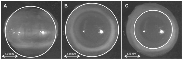
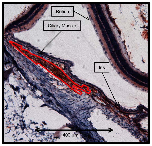
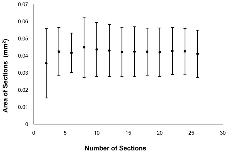
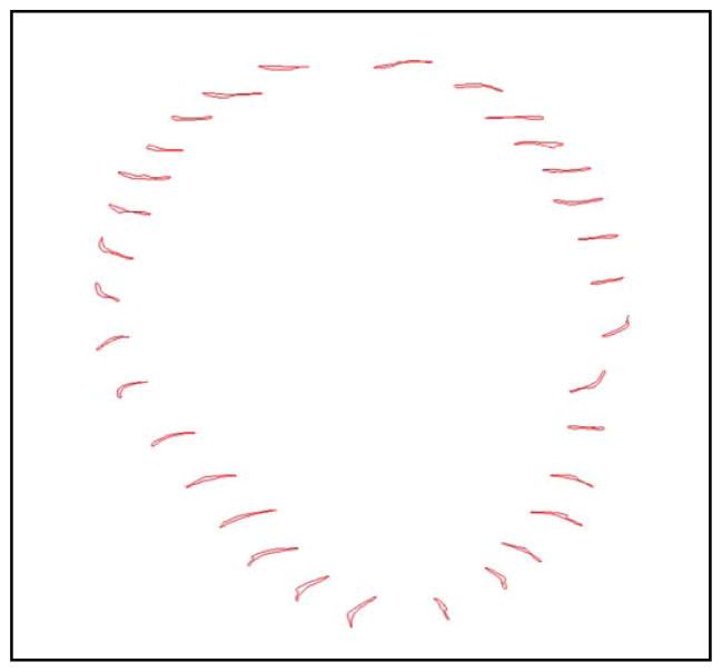
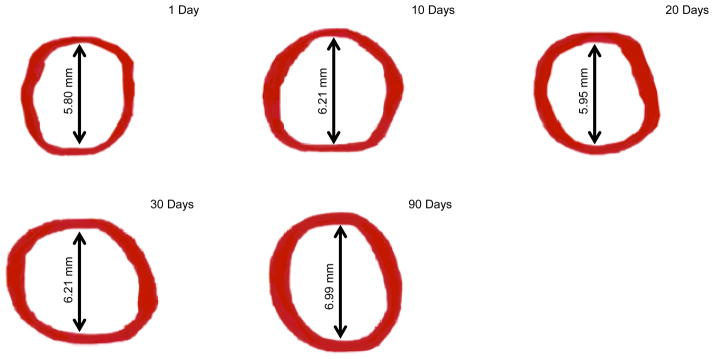
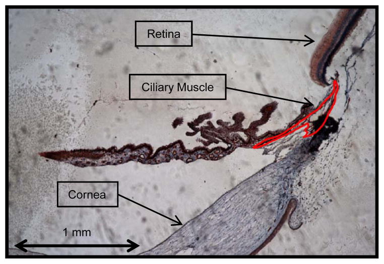
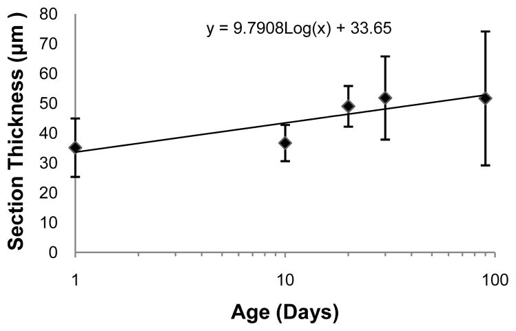
Methods: Six albino guinea pigs' eyes were collected at each of five ages (n = 30 eyes). Retinoscopy and photography were used to document refractive error, eye size, and eye shape. Serial sections through the excised eyes were made and then labeled with an α-smooth muscle actin antibody. The ciliary muscle was then visualized with an Olympus BX51 microscope, reconstructed with Stereo Investigator (MBF Bioscience), and analyzed using Neurolucida Explorer (MBF Bioscience). Full (using all sections) and partial (using a subset of sections) reconstruction methods were used to determine CMV.
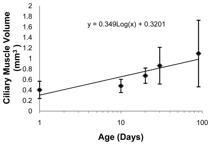
Results: There was no significant difference between the full and partial volume determination methods (p = 0.86). The mean (±SD) CMV of the 1-, 10-, 20-, 30-, and 90-day-old eyes was 0.40 (±0.16) mm, 0.48 (±0.13) mm, 0.67 (±0.15) mm, 0.86 (±0.35) mm, and 1.09 (±0.63) mm, respectively. Ciliary muscle volume was significantly correlated with log age (p = 0.001), ocular length (p = 0.003), limbal circumference (p = 0.01), and equatorial diameter (p = 0.003). It was not correlated with refractive error (p = 0.73) or eye shape (p = 0.60). Multivariate regression determined that biometric variables were not significantly associated with CMV after adjustment for age.
Conclusions: Three-dimensional reconstruction was an effective means of determining CMV. These data provide evidence that ciliary muscle growth occurs with age in tandem with eye size in normal albino guinea pigs. Additional work is needed to determine the relationship between CMV and abnormal ocular growth.
Figures

Similar articles
-
Morphological ciliary muscle changes associated with form deprivation-induced myopia.Exp Eye Res. 2020 Apr;193:107963. doi: 10.1016/j.exer.2020.107963. Epub 2020 Feb 8. Exp Eye Res. 2020. PMID: 32045599 Free PMC article.
-
Ciliary Muscle Cell Changes During Guinea Pig Development.Invest Ophthalmol Vis Sci. 2015 Dec;56(13):7691-6. doi: 10.1167/iovs.15-17927. Invest Ophthalmol Vis Sci. 2015. PMID: 26641547 Free PMC article.
-
Study of the establishment of a guinea pig model of ocular accommodative spasm by carbachol eye drops.Int Ophthalmol. 2024 Nov 11;44(1):425. doi: 10.1007/s10792-024-03334-z. Int Ophthalmol. 2024. PMID: 39527328
-
Quantification of age-related and per diopter accommodative changes of the lens and ciliary muscle in the emmetropic human eye.Invest Ophthalmol Vis Sci. 2013 Feb 7;54(2):1095-105. doi: 10.1167/iovs.12-10619. Invest Ophthalmol Vis Sci. 2013. PMID: 23287789 Free PMC article.
-
[Role of ciliary muscle in ocular physiology and disease].Vestn Oftalmol. 1999 Mar-Apr;115(2):13-5. Vestn Oftalmol. 1999. PMID: 10377866 Review. Russian.
Cited by
-
Morphological ciliary muscle changes associated with form deprivation-induced myopia.Exp Eye Res. 2020 Apr;193:107963. doi: 10.1016/j.exer.2020.107963. Epub 2020 Feb 8. Exp Eye Res. 2020. PMID: 32045599 Free PMC article.
-
Co-existence of myopia and amblyopia in a guinea pig model with monocular form deprivation.Ann Transl Med. 2021 Jan;9(2):110. doi: 10.21037/atm-20-5433. Ann Transl Med. 2021. PMID: 33569412 Free PMC article.
-
Optic nerve head and intraocular pressure in the guinea pig eye.Exp Eye Res. 2016 May;146:7-16. doi: 10.1016/j.exer.2015.12.007. Epub 2015 Dec 15. Exp Eye Res. 2016. PMID: 26698659 Free PMC article.
-
Ciliary Muscle Cell Changes During Guinea Pig Development.Invest Ophthalmol Vis Sci. 2015 Dec;56(13):7691-6. doi: 10.1167/iovs.15-17927. Invest Ophthalmol Vis Sci. 2015. PMID: 26641547 Free PMC article.
-
Study of the establishment of a guinea pig model of ocular accommodative spasm by carbachol eye drops.Int Ophthalmol. 2024 Nov 11;44(1):425. doi: 10.1007/s10792-024-03334-z. Int Ophthalmol. 2024. PMID: 39527328
References
-
- Lu F, Zhou X, Jiang L, Fu Y, Lai X, Xie R, Qu J. Axial myopia induced by hyperopic defocus in guinea pigs: A detailed assessment on susceptibility and recovery. Exp Eye Res. 2009;89:101–8. - PubMed
-
- Smith EL, 3rd, Hung LF. The role of optical defocus in regulating refractive development in infant monkeys. Vision Res. 1999;39:1415–35. - PubMed
-
- Howlett MH, McFadden SA. Spectacle lens compensation in the pigmented guinea pig. Vision Res. 2009;49:219–27. - PubMed
-
- Lawrence MS, Azar DT. Myopia and models and mechanisms of refractive error control. Ophthalmol Clin North Am. 2002;15:127–33. - PubMed
Publication types
MeSH terms
Substances
Grants and funding
LinkOut - more resources
Full Text Sources
Other Literature Sources
Medical
