. 2014 Jun 5;9(6):e99013.
doi: 10.1371/journal.pone.0099013. eCollection 2014.

Structure-function relationship of SW-AT-1, a serpin-type protease inhibitor in silkworm

Affiliations

Structure-function relationship of SW-AT-1, a serpin-type protease inhibitor in silkworm

Cheng Liu et al. PLoS One. .

Abstract

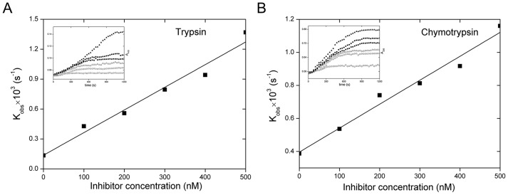
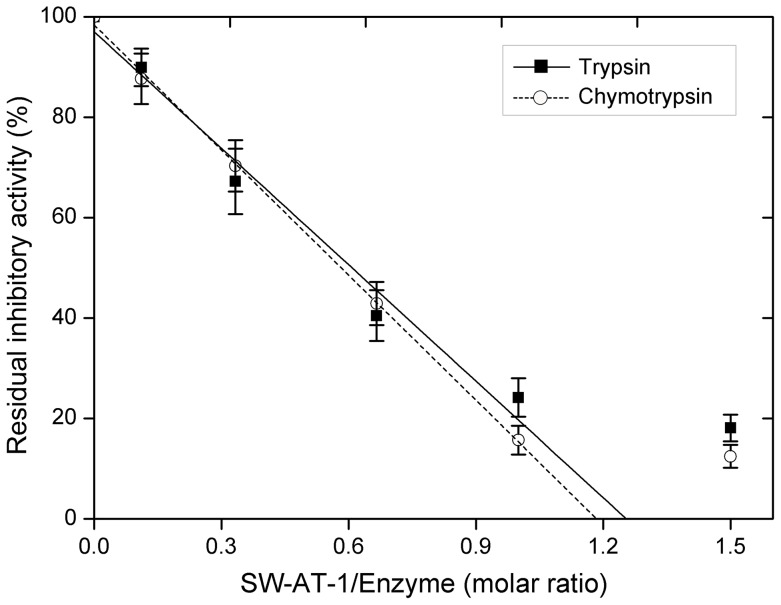
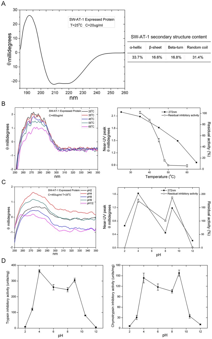
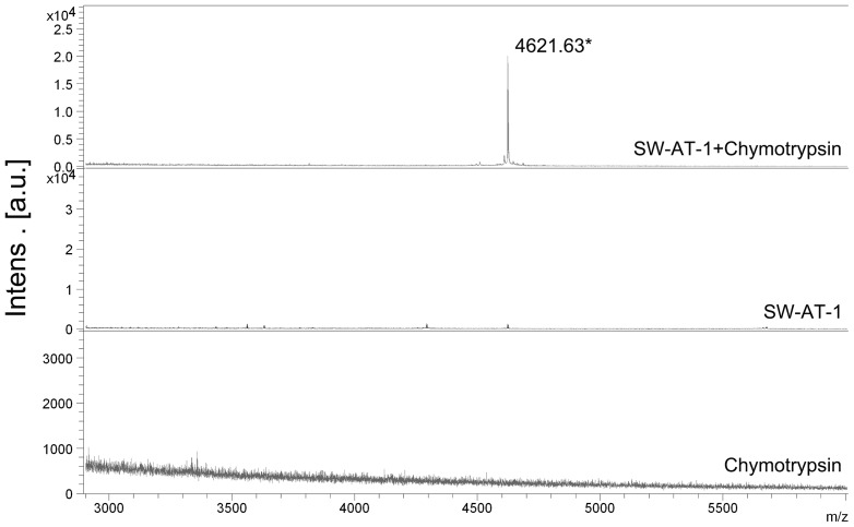
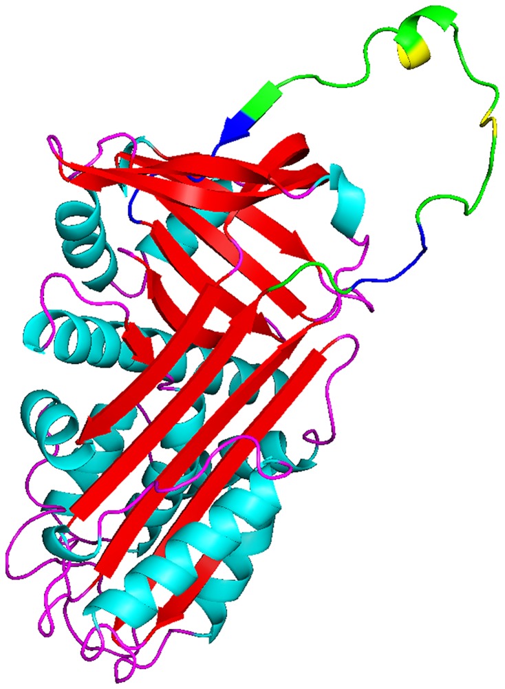
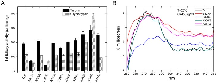
Although SW-AT-1, a serpin-type trypsin inhibitor from silkworm (Bombyx mori), was identified in previous study, its structure-function relationship has not been studied. In this study, SW-AT-1 was cloned from the body wall of silkworm and expressed in E. coli. rSW-AT-1 inhibited both trypsin and chymotrypsin in a concentration-dependent manner. The association rate constant for rSW-AT-1 and trypsin is 1.31×10⁻⁵ M-1s-1, for rSW-AT-1 and chymotrpsin is 2.85×10⁻⁶ M-1s-1. Circular dichroism (CD) assay showed 33% α-helices, 16% β-sheets, 17% turns, and 31% random coils in the secondary structure of the protein. Enzymatic and CD analysis indicated that rSW-AT-1 was stable at wide pH range between 4-10, and exhibited the highest activity at weakly acidic or alkaline condition. The predicted three-dimensional structure of SW-AT-1 by PyMOL (v1.4) revealed a deductive reactive centre loop (RCL) near the C-terminus, which was extended from the body of the molecule. In addition to trypsin cleavage site in RCL, matrix-assisted laser desorption ionization time of flight mass spectrometry indicated that the chymotrypsin cleavage site of SW-AT-1 was between F336 and T337 in RCL. Directed mutagenesis indicated that both the N- and C-terminal sides of RCL have effects on the activity, and G327 and E329 played an important role in the proper folding of RCL. The physiological role of SW-AT-1 in the defense responses of silkworm were also discussed.


Conflict of interest statement

Competing Interests: The authors have declared that no competing interests exit.

Figures

Figure 1
Figure 1. SDS-PAGE of recombinant His-tagged SW-AT-1.
Lane M, molecular size markers; lane 1, pET-28a vacant vector; lane 2, total protein extracts from E. coli after IPTG-induction; lane 3, total protein extracts from E. coli without IPTG-induction; lane 4, eluted fraction after affinity chromatography. Arrowhead indicated purified rSW-AT-1.
Figure 2
Figure 2. Determination of association rate constant.
(A) Trypsin (8 nM) was added to a mixture of 760 µM BAPNA and SW-AT-1 at 0 (□), 100 (○), 200 (Δ), 300 (▪), 400 (•) and 500(▴) nM. (B) Chymotrypsin (8 nM) was added to a mixture of 250 µM BTEE and SW-AT-1 at 0 (□), 100 (○), 200 (Δ), 300 (▪), 400 (•) and 500(▴) nM. The progress of enzyme inactivation (inset) was followed by measuring absorbance at 410 nm and 256nm on a microplate reader. Pseudo-first-order rate constants of inhibition (kobs) were plotted as a function of SW-AT-1 concentration ([I]).
Figure 3
Figure 3. Stoichiometry of inhibition.
Trypsin and chymotrypsin were incubated respectively with different concentrations of SW-AT-1 at 25°C for 20 min in the appropriate reaction buffer. Residual enzyme activity was measured by adding the appropriate substrate and determining the reaction velocity. The stoichiometry of inhibition was determined by using linear regression to extrapolate to that initial inhibitor/enzyme ratio resulting in complete inhibition of the enzyme.
Figure 4
Figure 4. Characterization of SW-AT-1.
(A) far-UV CD spectra of rSW-AT-1(left panel) and secondary sructure content (right panel); (B) near-UV CD spectra of rSW-AT-1 at different temperatures (left panel) and maximum aborbance 272-nm and trypsin inhibitory activity at different temperatures (right panel); (C) near-UV CD spectra of rSW-AT-1 at different pHs (left panel) and maximum aborbance at 272-nm and trypsin inhibitory activity at different pHs (right panel); (D) Optimal pH of rSW-AT-1 inhibitory activity for trypsin (left panel) and chymotrypsin (right panel). Bar indicates standard deviation from triplicate determination. P≤0.05 was considered statistically significant.
Figure 5
Figure 5. Cleavage site determination of SW-AT-1.
Chymotrypsin, rSW-AT-1, and the reaction mixture of SW-AT-1+chymotrypsin were directly analyzed by MALDI-TOF mass spectrometry. A representative strong single-accumulation spectrum is presented with the mass values on top of the MH+ peaks at 4623.613 Da. The spectrum was subjected to noise removal and calibrated with an external standard of bovine serum albumin.
Figure 6
Figure 6. The predicted three-dimensional structure of SW-AT-1.
SERPIN Domains was depicted as green, reactive site was depicted as yellow, and mutant sites were depicted as blue. The N and C termini were labeled as N and C, respectively. The structrue was generated using Open-Source PyMOL (v1.4), using Manduca sexta Serpin-protease 1K complex (1SEK) as the template.
Figure 7
Figure 7. Effects of N- and C-terminal side regions of RCL on the activity of SW-AT-1.
(A) Antitrypsin and antichymotrypsin activity. Data analysis was performed using the t-test function (P≤0.05). (B) near-UV CD spectra of WT and mutants.

Similar articles

Cited by

References

    1. Carrell RW, Pemberton PA, Boswell DR (1987) The Serpins: Evolution and adaptation in a family of protease Inhibitors. Cold Spring Harb Symp Quant Biol 52: 527–535. - PubMed
    1. Rau JC, Beaulieu LM, Huntington JA, Church FC (2007) Serpins in thrombosis, hemostasis and fibrinolysis. J Thromb Haemost 5 Suppl 1: 102–15. - PMC - PubMed
    1. Grigoryev SA, Woodcock CL (1998) Chromatin structure in granulocytes. A link between tight compaction and accumulation of a heterochromatin-associated protein (MENT). J Biol Chem 273: 3082–3089. - PubMed
    1. Irving JA, Shushanov SS, Pike RN, Popova EY, Bromme D, et al. (2002) Inhibitory activity of a heterochromatin-associated serpin (MENT) against papain-like cysteine proteinases affects chromatin structure and blocks cell proliferation. J Biol Chem 277: 13192–13201. - PubMed
    1. Ligoxygakis P, Roth S, Reichhart JM (2003) A serpin regulates dorsalventral axis formation in the Drosophila embryo. Curr Biol 13: 2097–2102. - PubMed

Publication types

MeSH terms