

Review
doi: 10.1259/bjr.20140324.
Epub 2014 Jun 5.
Non-cutaneous melanoma: is there a role for (18)F-FDG PET-CT?
Affiliations
- PMID: 24901893
- PMCID: PMC4112397
- DOI: 10.1259/bjr.20140324
Item in Clipboard
Review
Non-cutaneous melanoma: is there a role for (18)F-FDG PET-CT?
Br J Radiol.
2014 Aug.
Abstract
Non-cutaneous melanomas (NCM) are diverse and relatively uncommon. They often differ from cutaneous melanomas in their epidemiology, genetic profile and biological behaviour. Despite the growing body of evidence regarding the utility of positron emission tomography (PET)/CT in cutaneous melanoma, the data on its use in NCM are scarce. In this review, we will summarize the existing literature and present cases from our experience with NCM to illustrate current knowledge on the potential role and limitations of fluorine-18 fludeoxyglucose PET/CT in NCM.
Figures
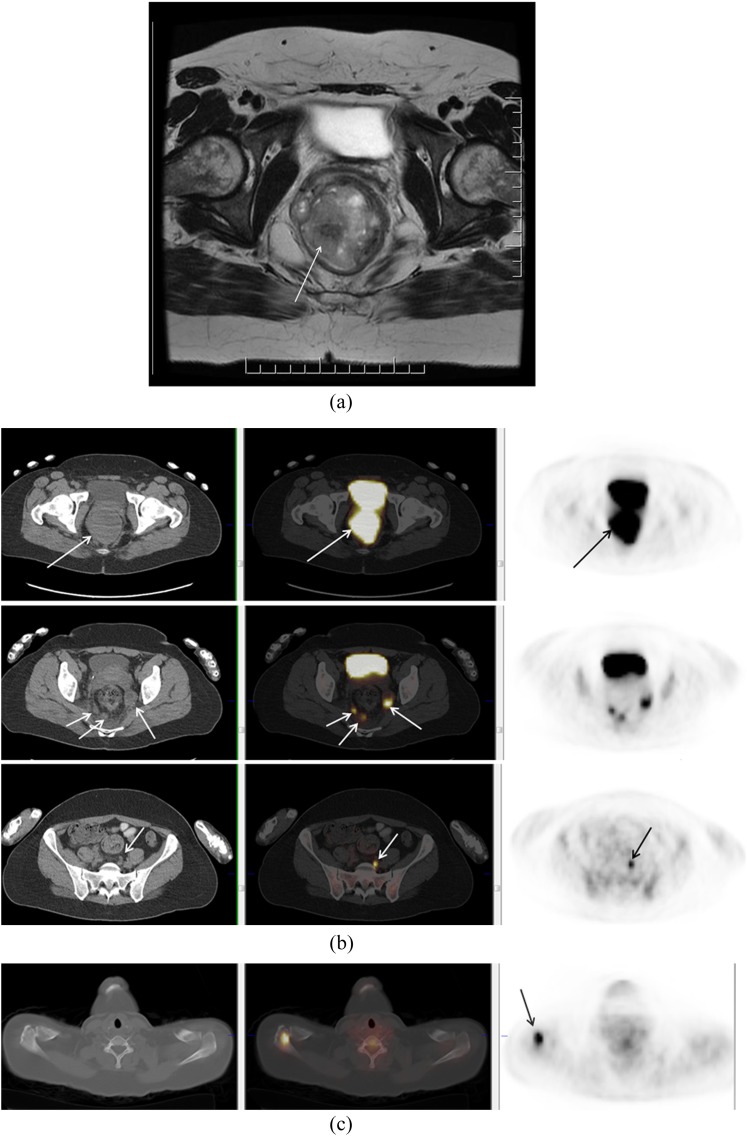
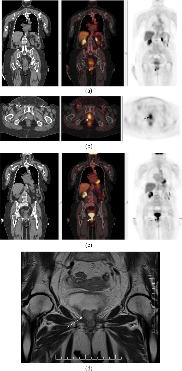
A 37-year-old female who presented with rectal bleeding. Biopsy of the rectal mass showed mucosal melanoma. A large tumour was identified on MRI (a). On fluorine-18 fludeoxyglucose positron emission tomography (PET)/CT, primary tumour and multiple nodal metastases in mesorectal space and left common iliac chain were identified (b). PET/CT also demonstrated a Segment 7 liver metastasis and a deposit in the right acromion (c).
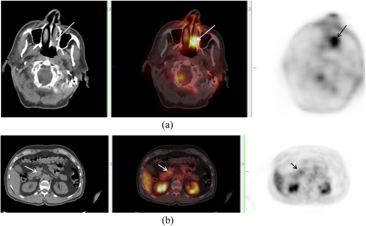
A 70-year-old male with left nasal melanoma (arrows) (a). Positron emission tomography/CT showed uptake in a mass at the head of the pancreas (arrows) (b). Biopsy confirmed metastatic deposit from nasal melanoma.
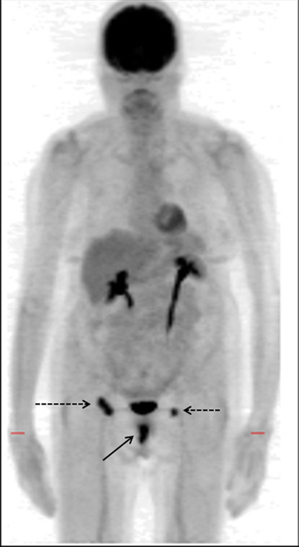
A 79-year-old female with vaginal melanoma (solid arrow). Positron emission tomography demonstrated uptake in primary lesion and bilateral inguinal nodes (dotted arrows).
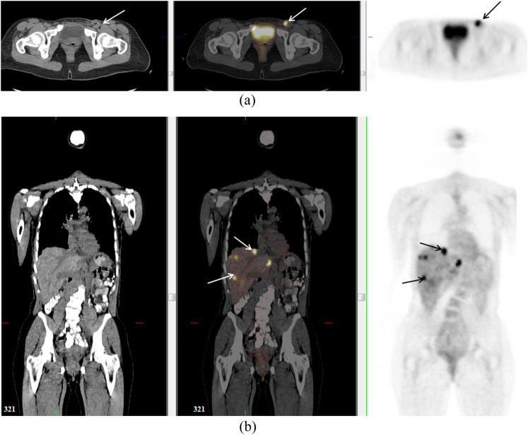
A 48-year-old female with vaginal melanoma. Positron emission tomography (PET)/CT confirmed uptake in left inguinal node (arrow) (a) but also detected previously unsuspected lesions in the liver (arrows) (b). These were not visualized on staging CT prior to PET but were seen on contrast-enhanced CT 4 months later (not shown).
A 47-year-old female with uveal melanoma. Coronal positron emission tomography-CT image demonstrated multiple fluorine-18 fludeoxyglucose avid bilateral hilar nodes. The pattern raised suspicion for an inflammatory aetiology, such as sarcoidosis, and transbronchial biopsy was performed. This was positive for non-necrotizing granulomatous inflammation.
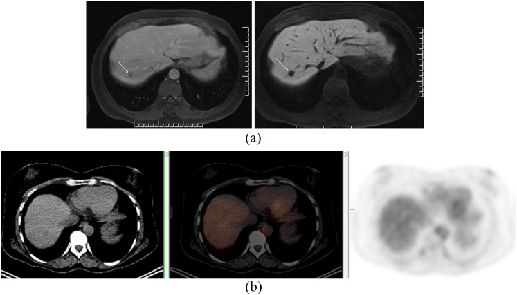
A 54-year-old female with uveal melanoma. Positron emission tomography (PET)/CT was negative; however, MR showed hepatic deposits. Gadolinium ethoxybenzyl dimeglumine (Primovist®; Bayer-Schering, Berlin, Germany)–enhanced MR (a) in the portal venous phase (left image) and in the hepatobiliary phase (right image) showing metastasis in Segment 7 (arrow). PET-CT at that level shows no abnormality (b).
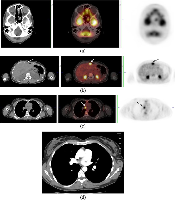
A 33-year-old female with melanoma from unknown primary. Positron emission tomography (PET)/CT demonstrated multiple deposits including in the sphenoid sinus (a), subcutaneous fat (not shown) and anterior to the left lobe of the liver (b). PET also demonstrated uptake of fluorine-18 fludeoxyglucose at the level of the right pulmonary artery (c), with no corresponding abnormality on concurrent contrast-enhanced CT chest (d). Follow-up CT 2 months later (not shown) confirmed a soft-tissue mass at that location.
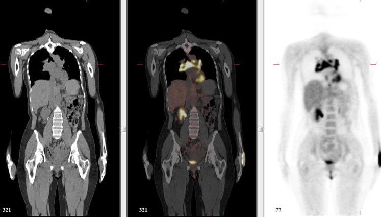
A 75-year-old female who had a previous resection of vaginal melanoma. She presented with right vulvar nodule, and a positron emission tomography (PET)/CT was ordered to assess for distant disease prior to local resection. Coronal PET image shows fluorine-18 fludeoxyglucose avid nodules in bilateral vulva (a) and an infiltrative soft-tissue tumour extending along the urethra to the bladder base (b, c) (axial and coronal PET/CT images through the urethra) (arrows). Coronal T2 weighted MR image confirmed intermediate signal intensity mass extending to bladder base (arrow) (d). The patient was deemed unsuitable for resection and received systemic therapy.
References
-
- Reinhardt MJ, Joe AY, Jaeger U, Huber A, Matthies A, Bucerius J, et al. Diagnostic performance of whole body dual modality 18F-FDG PET/CT imaging for N- and M-staging of malignant melanoma: experience with 250 consecutive patients. J Clin Oncol 2006; 24: 1178–87. doi: 10.1200/JCO.2005.03.5634 - DOI - PubMed
Publication types
MeSH terms
Substances
LinkOut - more resources
Full Text Sources
Other Literature Sources
Medical
