. 2014 Jun 24;53(24):4004-14.
doi: 10.1021/bi500072v. Epub 2014 Jun 13.

The ubiquitin-conjugating enzyme, UbcM2, is restricted to monoubiquitylation by a two-fold mechanism that involves backside residues of E2 and Lys48 of ubiquitin

Affiliations

The ubiquitin-conjugating enzyme, UbcM2, is restricted to monoubiquitylation by a two-fold mechanism that involves backside residues of E2 and Lys48 of ubiquitin

Linda Nguyen et al. Biochemistry. .

Abstract

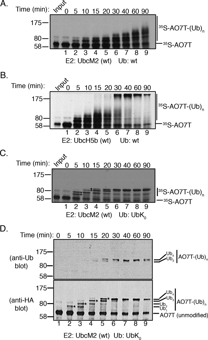
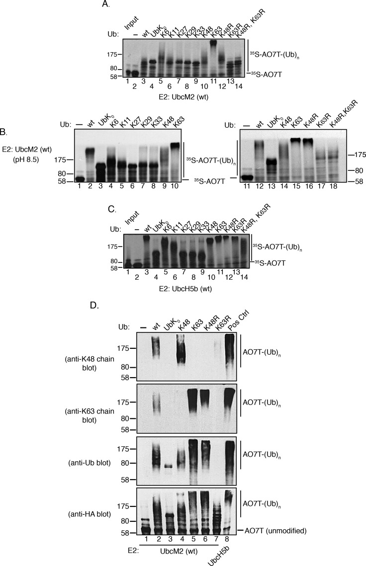
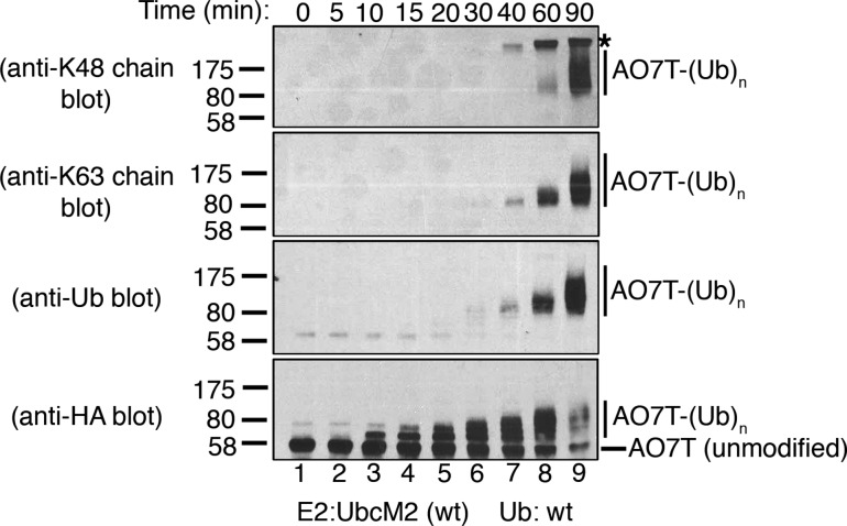
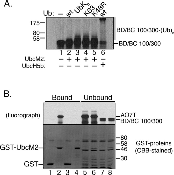
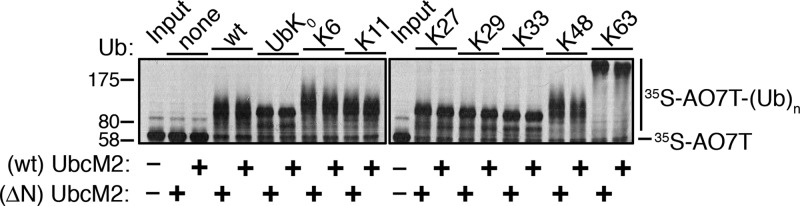
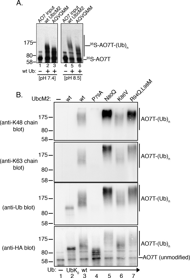
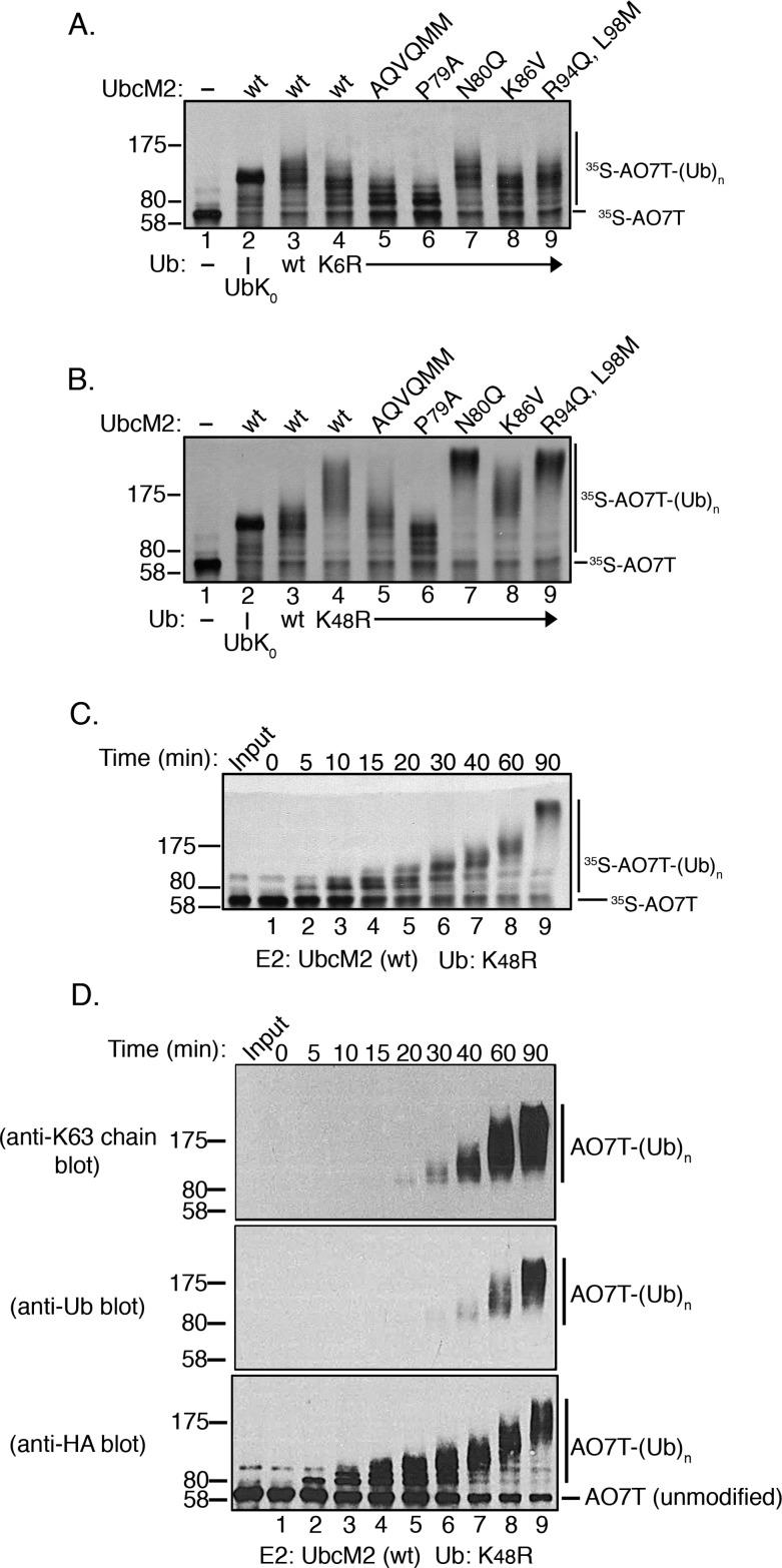
Proteins can be modified on lysines (K) with a single ubiquitin (Ub) or with polymers of Ub (polyUb). These different configurations and their respective topologies are primary factors for determining whether substrates are targeted to the proteasome for degradation or directed to nonproteolytic outcomes. We report here on the intrinsic ubiquitylation properties of UbcM2 (UBE2E3/UbcH9), a conserved Ub-conjugating enzyme linked to cell proliferation, development, and the cellular antioxidant defense system. Using a fully recombinant ubiquitylation assay, we show that UbcM2 is severely limited in its ability to synthesize polyUb chains with wild-type Ub. Restriction to monoubiquitylation is governed by multiple residues on the backside of the enzyme, far removed from its active site, and by lysine 48 of Ub. UbcM2 with mutated backside residues can synthesize K63-linked polyUb chains and to a lesser extent K6- and K48-linked chains. Additionally, we identified a single residue on the backside of the enzyme that promotes monoubiquitylation. Together, these findings reveal that a combination of noncatalytic residues within the Ubc catalytic core domain of UbcM2 as well as a lysine(s) within Ub can relegate a Ub-conjugating enzyme to monoubiquitylate its cognate targets despite having the latent capacity to construct polyUb chains. The two-fold mechanism for restricting activity to monoubiquitylation provides added insurance that UbcM2 will not build polyUb chains on its substrates, even under conditions of high local Ub concentrations.


Figures

Figure 1
Figure 1
UbcM2 has a severely limited capacity to synthesize polyUb chains on AO7T with wt Ub. (A) Fully recombinant in vitro ubiquitylation assays containing recombinant E1, H6T-UbcM2, wt Ub, energy, and [35S]AO7T (produced in a bacterial TNT expression system) were incubated at 37 °C for the indicated times. Reaction mixtures were solubilized and processed for SDS–PAGE and fluorography. (B) Same as panel A except testing H6T-UbcH5b. For panels A and B, [35S]AO7T input was loaded to the left of lane 1 and the migration of molecular weight markers is denoted on the left. Unmodified [35S]AO7T is marked on the right of each fluorograph with a hashmark and Ub-modified [35S]AO7T [[35S]AO7T-(Ub)n] with a vertical line. (C) Assay similar to that in panel A except wt Ub has been replaced with lysine-less Ub (UbK0). Distinct bands representing the attachment of one, two, three, or four monoUbK0 molecules to AO7T are denoted with asterisks between lanes 2 and 3 and lanes 3 and 4. (D) Assay similar to that in panel C except that HA-AO7T is not radiolabeled with [35S]Met/Cys but rather reaction products are visualized by either anti-Ub (top) or anti-HA Western blotting (bottom). The number of UbK0 molecules conjugated to HA-AO7T is indicated to the right of each blot. The sensitivity of the anti-Ub antibody is such that only HA-AO7T modified with either three or four UbK0 molecules is detected. All experiments were repeated at least three independent times.
Figure 2
Figure 2
UbcM2 can synthesize K63-linked polyUb chains. (A) H6T-UbcM2 in vitro ubiquitylation assays conducted at pH 7.4 with either no Ub (lane 2), wt Ub (lane 3), lysine-less Ub (UbK0, lane 4), Ub variants in which only the indicated lysine is intact and the six other lysines have been mutated to arginine (lanes 5–11), or variants in which only the indicated lysine has been mutated to arginine (lanes 12–14). Unmodified [35S]AO7T is marked on the right with a hashmark, and Ub-modified [35S]AO7 [[35S]AO7T-(Ub)n] is marked with a vertical line. The migration of molecular weight markers is denoted at the left. (B) Same as panel A except reactions were conducted at pH 8.5 and samples were run in parallel on two gels. (C) Same as panel A except testing H6T-UbcH5b. (D) Recombinant ubiquitylation assays using nonradiolabeled HA-AO7T were incubated for 90 min with the indicated variants of Ub. Reaction products were analyzed by Western blotting with the antibody listed to the left of each blot. As a positive control for the synthesis of K48- and K63-linked polyUb chains, UbcM2 was replaced with UbcH5b (lane 8, Pos Ctrl). All experiments were repeated a minimum of three independent times.
Figure 3
Figure 3
UbcM2 can synthesize K48- and K63-linked polyUb chains on AO7T. Recombinant ubiquitylation assays using nonradiolabeled HA-AO7T and wt Ub were incubated for the indicated times and solubilized, and the reaction products were analyzed by Western blotting. Unmodified AO7T is marked to the right of the anti-HA blot with a hashmark, and Ub-modified AO7T [AO7T-(Ub)n] is marked with a vertical line to the right of each blot. The migration of molecular weight markers and the antibodies used are denoted at the left. The asterisk denotes a nonspecific band detected by the anti-K48 linkage antibody. This band is derived from either a protein present in the energy-regenerating system or the PUREXPRESS bacterial lysate used to synthesize HA-AO7T. This band is detected at the 40, 60, and 90 min time points when AO7T is absent (i.e., unprogrammed PUREXPRESS bacterial lysate is used in the ubiquitylation reaction) (data not shown). Experiments were repeated a minimum of three independent times.
Figure 4
Figure 4
Affinity of UbcM2 for a proxy substrate that correlates with the capacity to attach polyUb chains. (A) In vitro ubiquitylation assay with H6T-UbcM2 and a 35S-labeled fusion of BARD1/BRCA1 100/300 (BD/BC 100/300) consisting of residues 26–126 of BARD1 fused to residues 1–304 of BRCA1. The Ub variant used in each reaction is indicated at the top of the fluorograph. In lane 6, H6T-UbcH5b was used in place of H6T-UbcM2. The migrations of unmodified BD/BC 100/300 and the Ub-modified fusion protein [BD/BC 100/300-(Ub)n] are indicated at the right, and the migration of molecular weight markers is denoted at the left. (B) Parallel GST fusion protein pull downs of 35S-labeled AO7T or 35S-labeled BD/BC 100/300. Fractions of bead-bound (75% of total) and unbound (37.5% of total) proteins were resolved by SDS–PAGE. 35S-labeled proteins were visualized by fluorography (top) and GST proteins by CBB staining (bottom). Assays were repeated a minimum of three independent times.
Figure 5
Figure 5
Unique amino-terminal extension of UbcM2 does not dictate the in vitro Ub conjugating behavior of the enzyme when it is partnered with AO7T. In vitro ubiquitylation assays comparing the polyUb chain building capacity of wt and ΔN-UbcM2, a mutant lacking the N-terminal 58-amino acid extension. Ub variants included in each 90 min reaction are indicated above the panels. The migrations of unmodified ([35S]AO7T) and ubiquitylated AO7T [[35S]AO7T-(Ub)n] are marked at the right and molecular weight markers at the left. The assay was repeated a minimum of three independent times.
Figure 6
Figure 6
Select noncatalytic backside residues influence the capacity of UbcM2 to synthesize polyUb chains. (A) In vitro ubiquitylation assays conducted at pH 7.4 (left) or pH 8.5 (right) to compare the patterns of [35S]AO7T ubiquitylation between wt UbcM2 and AQVQMM, a six-residue substitution mutant of UbcM2. Reaction mixtures contained either no Ub (lanes 1 and 4) or wt Ub (lanes 2, 3, 5, and 6). (B) Recombinant ubiquitylation assays using nonradiolabeled HA-AO7T were incubated for 90 min and contained either no Ub (lane 1), UbK0 (lane 2), or wt Ub (lanes 3–7), and the indicated forms of UbcM2 (listed above blots). Reaction products were analyzed by Western blotting with the antibody listed at the left of each blot. The migrations of unmodified AO7T and ubiquitylated AO7T [AO7T-(Ub)n] are shown to the right of the blots. The migration of molecular weight markers is indicated to the left of each blot. All assays were repeated a minimum of three independent times.
Figure 7
Figure 7
K6 and K48 of Ub differentially influence the ubiquitylating activity of UbcM2. (A and B) The wild type or the indicated substitution mutants of UbcM2 were incubated with K6R Ub (A) or K48R Ub (B) for 90 min at pH 7.4 (lanes 4–9). Control reaction mixtures contained either no Ub (lane 1), UbK0 (lane 2), or wt Ub (lane 3) for comparison. Ubiquitylation of [35S]AO7T was analyzed by SDS–PAGE and fluorography. (C) Kinetic assay analyzing the ubiquitylation activity of wt UbcM2 using K48R Ub at pH 7.4. Samples were incubated at 37 °C for the indicated times, solubilized, and processed for SDS–PAGE and fluorography. For panels A–C, the migrations of unmodified ([35S]AO7T) and ubiquitylated AO7T [[35S]AO7T-(Ub)n] are marked on the right and molecular weight markers on the left. (D) Similar to panel C using nonradiolabeled HA-AO7T and analyzing reaction products by Western blotting with the indicated antibodies.

References

    1. Olsen S. K.; Lima C. D. (2013) Structure of a ubiquitin E1-E2 complex: Insights to E1-E2 thioester transfer. Mol. Cell 49, 884–896. - PMC - PubMed
    1. Ye Y.; Rape M. (2009) Building ubiquitin chains: E2 enzymes at work. Nat. Rev. Mol. Cell Biol. 10, 755–764. - PMC - PubMed
    1. Chau V.; Tobias J. W.; Bachmair A.; Marriott D.; Ecker D. J.; Gonda D. K.; Varshavsky A. (1989) A multiubiquitin chain is confined to specific lysine in a targeted short-lived protein. Science 243, 1576–1583. - PubMed
    1. Finley D.; Sadis S.; Monia B. P.; Boucher P.; Ecker D. J.; Crooke S. T.; Chau V. (1994) Inhibition of proteolysis and cell cycle progression in a multiubiquitination-deficient yeast mutant. Mol. Cell. Biol. 14, 5501–5509. - PMC - PubMed
    1. Thrower J. S.; Hoffman L.; Rechsteiner M.; Pickart C. M. (2000) Recognition of the polyubiquitin proteolytic signal. EMBO J. 19, 94–102. - PMC - PubMed

Publication types

MeSH terms

LinkOut - more resources