. 2014 Jun 5:15:169.
doi: 10.1186/1471-2105-15-169.

A semi-automated technique for labeling and counting of apoptosing retinal cells

Affiliations

A semi-automated technique for labeling and counting of apoptosing retinal cells

Mukhtar Bizrah et al. BMC Bioinformatics. .

Abstract

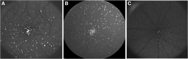
Background: Retinal ganglion cell (RGC) loss is one of the earliest and most important cellular changes in glaucoma. The DARC (Detection of Apoptosing Retinal Cells) technology enables in vivo real-time non-invasive imaging of single apoptosing retinal cells in animal models of glaucoma and Alzheimer's disease. To date, apoptosing RGCs imaged using DARC have been counted manually. This is time-consuming, labour-intensive, vulnerable to bias, and has considerable inter- and intra-operator variability.

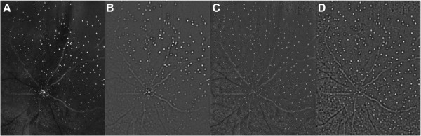
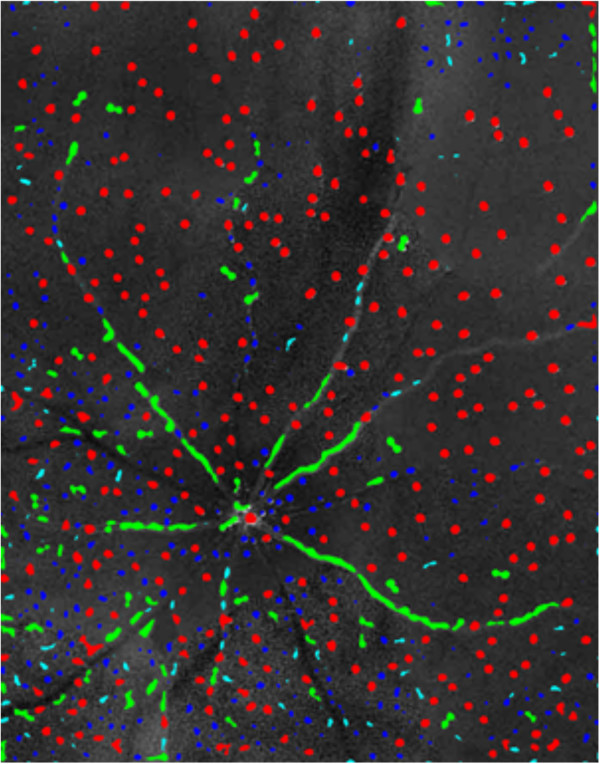
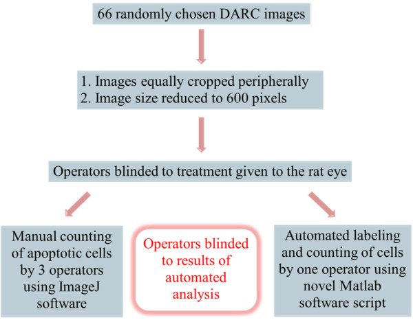
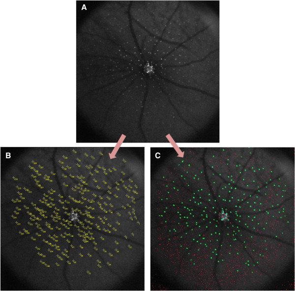
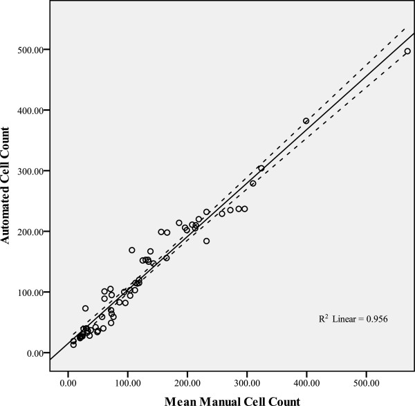
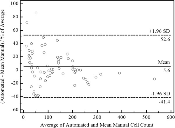
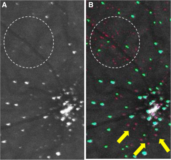
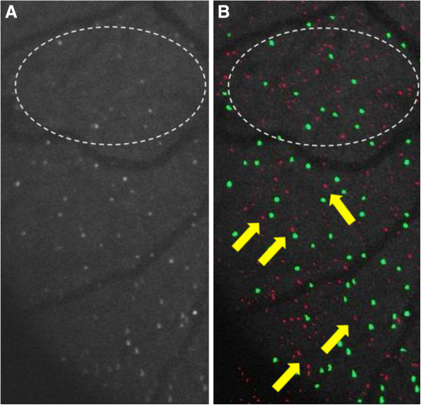
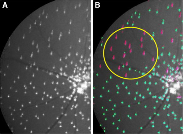
Results: A semi-automated algorithm was developed which enabled automated identification of apoptosing RGCs labeled with fluorescent Annexin-5 on DARC images. Automated analysis included a pre-processing stage involving local-luminance and local-contrast "gain control", a "blob analysis" step to differentiate between cells, vessels and noise, and a method to exclude non-cell structures using specific combined 'size' and 'aspect' ratio criteria. Apoptosing retinal cells were counted by 3 masked operators, generating 'Gold-standard' mean manual cell counts, and were also counted using the newly developed automated algorithm. Comparison between automated cell counts and the mean manual cell counts on 66 DARC images showed significant correlation between the two methods (Pearson's correlation coefficient 0.978 (p < 0.001), R Squared = 0.956. The Intraclass correlation coefficient was 0.986 (95% CI 0.977-0.991, p < 0.001), and Cronbach's alpha measure of consistency = 0.986, confirming excellent correlation and consistency. No significant difference (p = 0.922, 95% CI: -5.53 to 6.10) was detected between the cell counts of the two methods.

Conclusions: The novel automated algorithm enabled accurate quantification of apoptosing RGCs that is highly comparable to manual counting, and appears to minimise operator-bias, whilst being both fast and reproducible. This may prove to be a valuable method of quantifying apoptosing retinal cells, with particular relevance to translation in the clinic, where a Phase I clinical trial of DARC in glaucoma patients is due to start shortly.


Figures

Figure 1
Figure 1
Images A, B & C are examples of DARC images before undergoing manual or automated cell labeling.
Figure 2
Figure 2
The effects of pre-processing illustrated on a DARC image. (A) A raw DARC image. (B) The same image filtered with a Δ2Gt filter (C) A pre-processed version of (A) (corrected for local variation in luminance structure). (D) A Laplacian filtered version of (C). Compare (D) to (B) and note presence of additional image structure in (D).
Figure 3
Figure 3
An illustration of the DARC image shown in Figure 2d after undergoing thresholding and a novel ‘blob analysis’ stage to classify blobs as cells (red), vessels (green) or noise (blue).
Figure 4
Figure 4
Flowchart summarizing the protocol followed for the manual and automated analysis of the DARC images.
Figure 5
Figure 5
Example of a DARC image before and after undergoing manual and automated labeling. Image A represents a cropped DARC image before undergoing labeling. Image B represents the same DARC image after undergoing manual labeling using ImageJ® ‘multi-point selections’ tool, which marks and numbers each selected spot on the image. Image C represents the same DARC image after undergoing automated cell labeling using the novel Matlab® script. In Image C, structures identified as ‘cells’ have been labeled in green, whilst ‘non-cellular’ structures have been labeled in red.
Figure 6
Figure 6
Correlation between the Mean Manual Cell Counts and the Automated Cell Counts of the 66 DARC images. The continuous line is the best-fit line, and the adjacent dotted lines represent the 95% confidence intervals.
Figure 7
Figure 7
Bland-Altman Plot of Percent Difference of Automated Cell Counts from Mean Manual Cell Counts.
Figure 8
Figure 8
The first DARC image with cell count differences beyond the 95% limits of agreement.
Figure 9
Figure 9
The second DARC image with cell count differences beyond the 95% limits of agreement.
Figure 10
Figure 10
Example of a cropped and magnified section of a DARC before (A) and after (B) undergoing automated labeling. Structures identified by the automated algorithm as cells are shown as green spots, whereas spots identified by the algorithm as non-cellular structures are shown as pink spots.
Figure 11
Figure 11
Example of a cropped and magnified section of a DARC image before (A) and after (B) undergoing automated labeling.
Figure 12
Figure 12
Example of a cropped and magnified section of a DARC before (A) and after (B) undergoing automated labeling.

References

    1. Bagga H, Greenfield DS. Quantitative assessment of structural damage in eyes with localized visual field abnormalities. Am J Ophthalmol. 2004;137(5):797–805. doi: 10.1016/j.ajo.2003.11.060. - DOI - PubMed
    1. Quigley HA, Nickells RW, Kerrigan LA, Pease ME, Thibault DJ, Zack DJ. Retinal ganglion cell death in experimental glaucoma and after axotomy occurs by apoptosis. Invest Ophthalmol Vis Sci. 1995;36(5):774–786. - PubMed
    1. Bizrah MG, Guo L, Cordeiro MF. Glaucoma and Alzheimer’s disease in the elderly. Aging Health. 2011;7(5):719–733. doi: 10.2217/ahe.11.51. - DOI
    1. Friedlander RM. Apoptosis and caspases in neurodegenerative diseases. N Engl J Med. 2003;348(14):1365–1375. doi: 10.1056/NEJMra022366. - DOI - PubMed
    1. Berry MD, Boulton AA. Apoptosis and human neurodegenerative diseases. Prog Neuropsychopharmacol Biol Psychiatry. 2003;27(2):197–198. doi: 10.1016/S0278-5846(03)00015-0. - DOI - PubMed

Publication types