. 2014 Jul;24(7):842-51.
doi: 10.1038/cr.2014.74. Epub 2014 Jun 6.

Ribosome profiling reveals sequence-independent post-initiation pausing as a signature of translation

Affiliations

Ribosome profiling reveals sequence-independent post-initiation pausing as a signature of translation

Yan Han et al. Cell Res. 2014 Jul.

Abstract

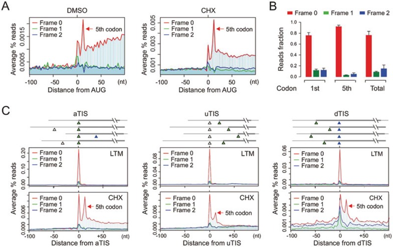
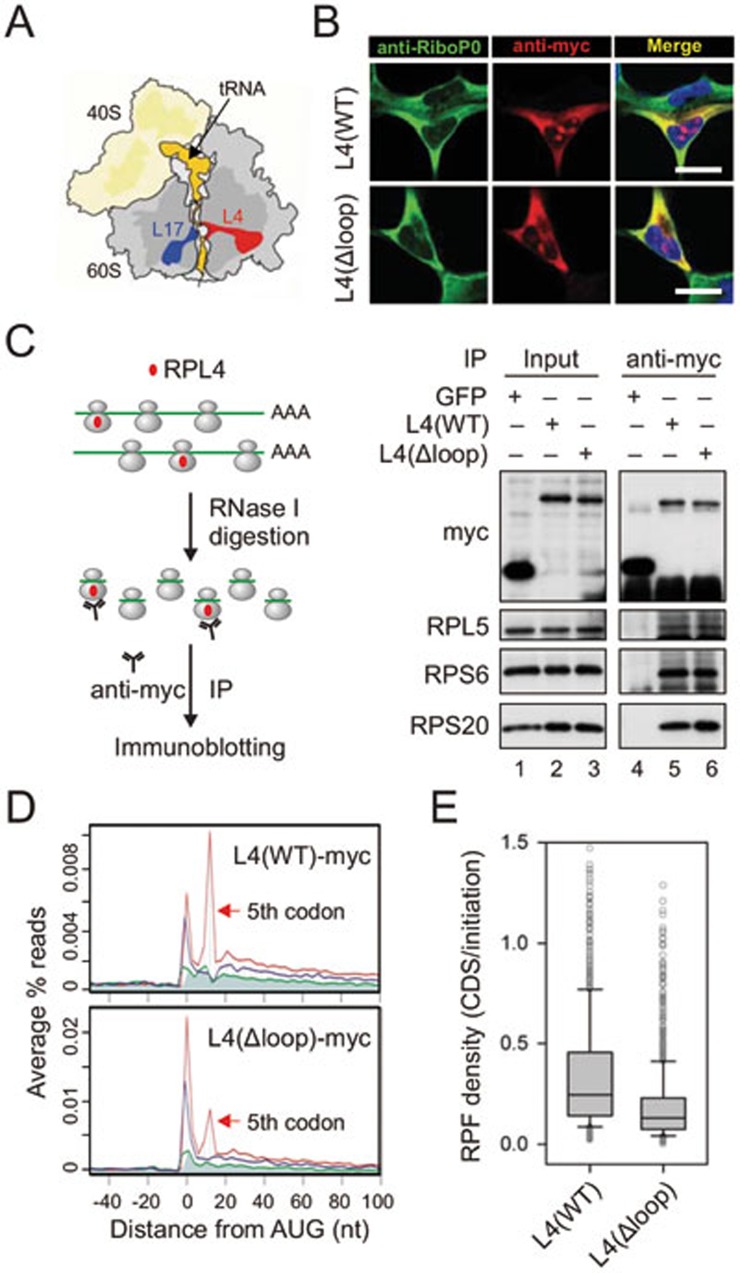
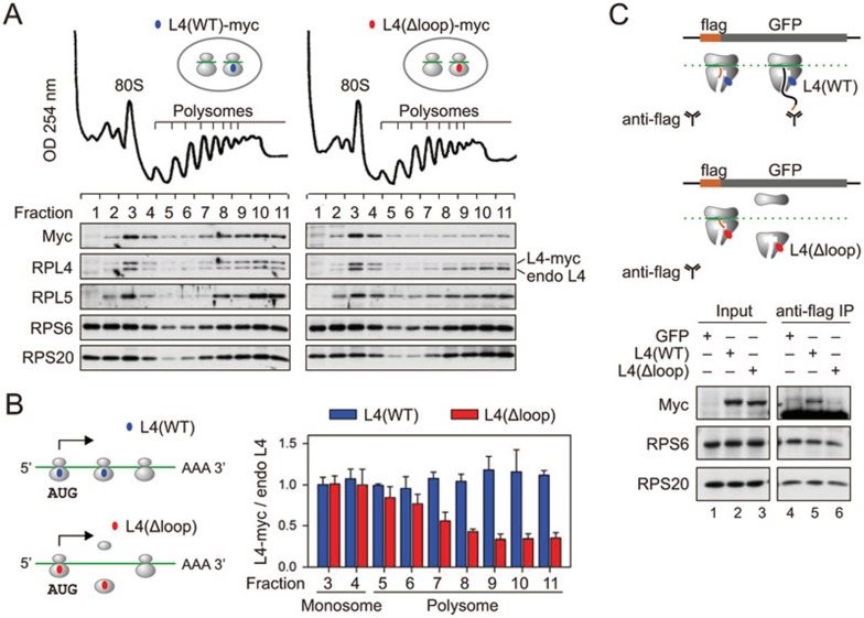
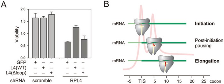
The journey of a newly synthesized polypeptide starts in the peptidyltransferase center of the ribosome, from where it traverses the exit tunnel. The interior of the ribosome exit tunnel is neither straight nor smooth. How the ribosome dynamics in vivo is influenced by the exit tunnel is poorly understood. Genome-wide ribosome profiling in mammalian cells reveals elevated ribosome density at the start codon and surprisingly the downstream 5th codon position as well. We found that the highly focused ribosomal pausing shortly after initiation is attributed to the geometry of the exit tunnel, as deletion of the loop region from ribosome protein L4 diminishes translational pausing at the 5th codon position. Unexpectedly, the ribosome variant undergoes translational abandonment shortly after initiation, suggesting that there exists an obligatory step between initiation and elongation commitment. We propose that the post-initiation pausing of ribosomes represents an inherent signature of the translation machinery to ensure productive translation.


Figures

Figure 1
Figure 1
Characterization of post-initiation pausing. (A) Metagene analysis of RPFs obtained from HEK293 cells treated with CHX (right) or DMSO control (left). All mapped reads are aligned at the annotated start codon AUG and stratified by frames. The read density at each nucleotide position is averaged using the P-site of RPFs. The red arrow indicates the 5th codon position. (B) Read fractions relative to the annotated reading frame are calculated for RPFs mapped to the 1st (AUG) codon, 5th codon, or the entire CDS. Data are shown as means ± SD, n = 4. (C) Metagene analysis of RPFs obtained from HEK293 cells treated with LTM (upper) or CHX (bottom). All mapped reads are aligned at the annotated start codon (aTIS, green triangle; left panel), upstream TIS (uTIS, white triangle; middle panel), or downstream TIS (dTIS, blue triangle; right panel). The read density at each nucleotide position is averaged using the P-site of RPFs. The red arrow indicates the 5th codon position.
Figure 2
Figure 2
Reduced post-initiation pausing for ribosomes bearing L4 mutant. (A) Schematic structure of ribosomes highlighting the exit tunnel. tRNA and the nascent chain are shown in orange, RPL4 in red, and RPL17 in blue. (B) Cellular localization of L4 wild type and Δloop mutant is examined in HEK293 cells using immunostaining. Scale bar, 10 μm. (C) Myc-tagged L4 is incorporated into ribosome complexes. Polysome fractions from transfected HEK293 cells were treated with RNase I prior to anti-myc IP. Both the input and the immunoprecipitates were blotted using antibodies as indicated. (D) Metagene analysis of RPFs obtained from purified ribosomes bearing L4 wild type (top) and Δloop mutant (bottom). All mapped reads are aligned at the annotated start codon AUG and stratified by frames. (E) Read density over CDS relative to the initiation site (1st - 5th) is shown in box plots.
Figure 3
Figure 3
Ribosomes bearing L4 mutant lead to abortive translation. (A) Polysome profiles of HEK293 cells expressing L4 wild type or loop mutant. The bottom panel shows the distribution of exogenous and endogenous ribosome proteins in ribosome fractions. (B) A simple model of translational abandonment for ribosomes bearing L4(Δloop) mutant. The right panel shows the quantification of myc-tagged L4 relative to the endogenous L4 in ribosome fractions as shown in A. Data are shown as means ± SD, n = 3. (C) Schematic for nascent chain IP assay using a HEK293 cell line expressing flag-GFP (top panel). Transfected HEK293 cells were lysed and treated with RNase I prior to anti-flag IP. Both the input and the immunoprecipitates were blotted for ribosomal proteins as indicated (bottom panel).
Figure 4
Figure 4
Physiological significance of post-initiation pausing. (A) HEK293 cells expressing GFP control, L4 wild type, or Δloop mutant were infected with lentiviruses encoding shRNA targeting endogenous L4 or scramble control. Cell viability was measured and quantified. Data are shown as means ± SD, n = 3. (B) A model for post-initiation pausing of ribosomes. After initiation, the nascent chain (red) traverses the exit tunnel (yellow). At the 5th codon position, the paused ribosome undergoes conformational changes, thereby facilitating elongation commitment by preventing nascent chain from off-route and abortive translation.

References

    1. Buchan JR, Stansfield I. Halting a cellular production line: responses to ribosomal pausing during translation. Biol Cell. 2007;99:475–487. - PubMed
    1. Kertesz M, Wan Y, Mazor E, et al. Genome-wide measurement of RNA secondary structure in yeast. Nature. 2010;467:103–107. - PMC - PubMed
    1. Plotkin JB, Kudla G. Synonymous but not the same: the causes and consequences of codon bias. Nat Rev Genet. 2011;12:32–42. - PMC - PubMed
    1. Kramer G, Boehringer D, Ban N, Bukau B. The ribosome as a platform for co-translational processing, folding and targeting of newly synthesized proteins. Nat Struct Mol Biol. 2009;16:589–597. - PubMed
    1. Farabaugh PJ. Translational frameshifting: implications for the mechanism of translational frame maintenance. Prog Nucleic Acid Res Mol Biol. 2000;64:131–170. - PubMed

Publication types