. 2014 Jun 6:14:184.
doi: 10.1186/1472-6882-14-184.

Effects of JSOG-6 on protection against bone loss in ovariectomized mice through regulation of osteoblast differentiation and osteoclast formation

Affiliations

Effects of JSOG-6 on protection against bone loss in ovariectomized mice through regulation of osteoblast differentiation and osteoclast formation

Hwa-Jin Chung et al. BMC Complement Altern Med. .

Abstract

Background: JSOG-6 is used as a traditional medicine to relieve the symptoms associated with inflammation, rheumatism, and osteoporosis in Korea. In the present study, we investigated the effects of JSOG-6 on bone loss prevention both in in vitro and in vivo as well as its underlying mechanism of action.

Methods: Protection against bone loss was assessed in an ovariectomized (OVX) mouse model. Bone microarchitecture was measured using a micro-computed tomography to detect the parameters of three-dimensional structure of a trabecular bone. Serum biomarkers were also evaluated in an OVX-induced model. Osteoclasts derived from mouse bone marrow cells (BMCs) and osteoblastic MC3T3-E1 cells were also employed to investigate the mechanism of action.

Results: Oral administration of JSOG-6 significantly increased the bone mineral density (BMD) of the femur in OVX mice in vivo. Especially, the reduced Tb.No (trabecular bone number) in the OVX group was significantly recovered by JSOG-6 treatment. The serum levels of alkaline phosphatase (ALP), osteocalcin, C-terminal telopeptide, and tartrate-resistant acid phosphatase, biomarkers of bone resorption, were significantly elevated in OVX mice, but JSOG-6 effectively inhibited the increase in OVX mice. JSOG-6 was also found to enhance the osteoblastic differentiation and maturation with the increase of the density and ALP activity, a marker of osteoblastic differentiation, as well as calcium deposition, a marker of osteoblastic maturation in MC3T3-E1 cells. The effects of JSOG-6 on osteoblastic differentiation were also associated in part with the increase of ALP and OPN mRNA expressions and the decrease of RANKL mRNA expression in MC3T3-E1 cells.

Conclusions: The findings demonstrate that JSOG-6 induced protection against bone loss in OVX mice, and its anti-osteoporotic property might be, in part, a function of the stimulation of osteoblast differentiation and the inhibition of osteoclast formation. These findings suggest that JSOG-6 might be an applicable therapeutic traditional medicine for the regulation of the osteoporotic response.


Figures

Figure 1
Figure 1
Effect of JSOG-6 on bone loss in OVX mice. (A) Change in body weight 12 weeks after ovarietomy. (B) Effect of JSOG-6 on bone 3D microCT image of the distal femur in OVX mice. (C) Effect of JSOG-6 on the bone morphometric parameters BMD, BV/TV (%), and Tb.No (1/mm) as analyzed with micro-CT SkyScan CTAN software. Data represent the mean ± S.D. (n = 8). *P < 0.01 indicates statistically significant differences from the OVX mice group.
Figure 2
Figure 2
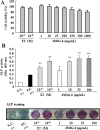
Effect of JSOG-6 on ALP activity in MC3T3-E1 cells. (A) Cell viability was measured by the MTT method as described in the Methods. (B) MC3T3-E1 cells (2 × 104 cells/mL) were incubated with JSOG-6 in the presence of ascorbic acid and β-glycerophosphate for 4 days. The ALP activity was corrected for the amount of protein. Data represent the mean ± S.D. (n = 3). *P < 0.05, **P < 0.01 indicates statistically significant differences from the control group. N.C., negative control; P.C., positive control (ascorbic acid + β-glycerophosphate).
Figure 3
Figure 3
Effect of JSOG-6 on the mineralization of MC3T3-E1 cells. (A) MC3T3-E1 cells (2 × 104 cells/mL) were incubated with JSOG-6 in the presence of ascorbic acid and β-glycerophosphate for 14 days. Mineralized nodule formation was assessed by Alizarin red S staining. Data represent the mean ± S.D. (n = 3). *P < 0.01 indicates statistically significant differences from the control group. (B) Representative microscopic observation of JSOG-6 on the formation of calcification nodules with staining Alizarin red S. N.C., negative control; P.C., positive control (ascorbic acid + β-glycerophosphate).
Figure 4
Figure 4
Effect of JSOG-6 on osteoblastic gene expression. MC3T3-E1 cells (2 × 104 cells/mL) were treated with the indicated concentrations of JSOG-6 for 48 h, and the mRNA levels of osteoblastic genes were examined using real-time PCR. The results are presented as a relative expression level compared to unstimulated cells and were normalized to β-actin. Data represent the mean ± S.D. (n = 3). *P < 0.05, **P < 0.01 indicates statistically significant differences from the control group.
Figure 5
Figure 5
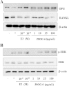
Effect of JSOG-6 on the protein levels of OPG, RANKL, and ERK in MC3T3-E1 cells. (A, B) MC3T3-E1 cells (2× 10⁴ cells/mL) were incubated for 48 h and then treated with JSOG-6 for 48 h. After incubation, total cell extracts were obtained and subjected to Western blot analysis as described in the Methods. Data were representative of three separate experiments. β-Actin was used as an internal standard.
Figure 6
Figure 6
Effect of JSOG-6 on RANKL-induced osteoclast differentiation. (A) Cell viability was measured by the MTT method as described in the Methods. (B) Bone marrow cells (1 × 104 cells/mL) were incubated with JSOG-6 in the presence of M-CSF (30 ng/mL) and RANKL (100 ng/mL) for 5 days. Osteoclastogenesis was confirmed by TRAP staining. Data represent the mean ± S.D. (n = 3). **P < 0.01 indicates statistically significant differences from the control group. (C) The expression of TRAP was determined by Western blot analysis as described in the Methods. Data are representative of three separated experiments. β-Actin was used as an internal standard.

References

    1. Alexander JM, Bab I, Fish S, Müller R, Uchiyama T, Gronowicz G, Nahounou M, Zhao Q, White DW, Chorev M, Gazit D, Rosenblatt M. Human parathyroid hormone 1–34 reverses bone loss in ovariectomized mice. J Bone Miner Res. 2001;161:665–673. - PubMed
    1. Raisz LG. Pathogenesis of osteoporosis: concepts, conflicts, and prospects. J Clin Invest. 2005;115:3318–3325. doi: 10.1172/JCI27071. - DOI - PMC - PubMed
    1. Takayanagi H. Inflammatory bone destruction and osteoimmunology. J Periodontal Res. 2005;40:287–293. doi: 10.1111/j.1600-0765.2005.00814.x. - DOI - PubMed
    1. Papachroni KK, Karatzas DN, Papavassiliou KA, Basdra EK, Papavassiliou AG. Mechanotransduction in osteoblast regulation and bone disease. Trends Mol Med. 2009;15:208–216. doi: 10.1016/j.molmed.2009.03.001. - DOI - PubMed
    1. Harada S, Rodan GA. Control of osteoblast function and regulation of bone mass. Nature. 2003;15:349–355. - PubMed

Publication types

MeSH terms