Review
. 2014 Jul;13(7):549-60.
doi: 10.1038/nrd4295. Epub 2014 Jun 6.

Muscarinic acetylcholine receptors: novel opportunities for drug development

Affiliations
Review

Muscarinic acetylcholine receptors: novel opportunities for drug development

Andrew C Kruse et al. Nat Rev Drug Discov. 2014 Jul.

Abstract

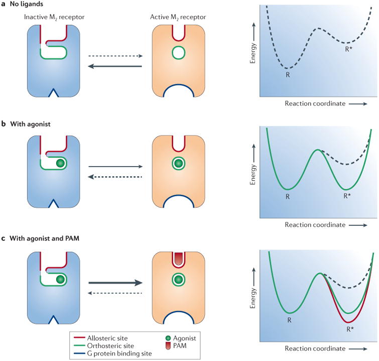
The muscarinic acetylcholine receptors are a subfamily of G protein-coupled receptors that regulate numerous fundamental functions of the central and peripheral nervous system. The past few years have witnessed unprecedented new insights into muscarinic receptor physiology, pharmacology and structure. These advances include the first structural views of muscarinic receptors in both inactive and active conformations, as well as a better understanding of the molecular underpinnings of muscarinic receptor regulation by allosteric modulators. These recent findings should facilitate the development of new muscarinic receptor subtype-selective ligands that could prove to be useful for the treatment of many severe pathophysiological conditions.


Figures

Figure 1
Figure 1. Modes of targeting mAChRs (GPCRs) by different classes of ligands
Orthosteric ligands (green) bind to the site recognized by the endogenous agonist (acetylcholine) for the receptor. Allosteric ligands (yellow) bind to a topographically distinct site. Bitopic ligands (blue) concomitantly interact with both orthosteric and allosteric sites. The key properties generally associated with each mode of receptor targeting are also indicated. GPCR, G protein-coupled receptor; mAChR, muscarinic acetylcholine receptor; SAR, structure–activity relationship.
Figure 2
Figure 2. Overall structure of the M2 and M3 receptors
The structures of the inactive, antagonist-bound M2 (blue) and M3 (yellow) receptors are highly similar to each other in overall fold. For the sake of clarity, only the M3 receptor ligand (tiotropium) is shown. The overall architecture of the M2 and M3 muscarinic acetylcholine receptors (mAChRs) is very similar to that of other biogenic amine G protein-coupled receptors (GPCRs), with similar orthosteric ligand binding sites (orange spheres).
Figure 3
Figure 3. Structure of the orthosteric mAChR ligand binding site
a | A cross-section through the M2 receptor structure reveals a large, solvent-accessible cavity extending through the receptor. The QNB ligand (orange spheres) is bound within the receptor transmembrane core (black, receptor protein; blue, receptor surface). b | The orthosteric ligand (antagonist)-binding sites for the M2 and M3 receptors are almost identical in both structure and sequence. Polar contacts (red dotted lines) between the receptor and bound antagonist (M2, QNB; M3, tiotropium) are identical for the two receptors. Residues are numbered according to the human M2 receptor sequence. mAChR, muscarinic acetylcholine receptor.
Figure 4
Figure 4. Activation and allosteric modulation of the M2 receptor
As shown in part a and part b, the intracellular tip of transmembrane domain 6 (TM6) rotates outwards in the active M2 receptor structure (orange) relative to the inactive state (blue). As shown in part c, the orthosteric binding site contracts upon M2 receptor activation, enclosing the agonist iperoxo (yellow) in a smaller binding site, as compared to the antagonist (QNB; green) binding cavity. Residues are numbered according to the human M2 receptor sequence. As shown in part d, LY2119620 (magenta), a muscarinic positive allosteric modulator, binds to the extracellular vestibule of the M2 receptor directly above the orthosteric agonist iperoxo (yellow). ICL2, intracellular loop 2.
Figure 5
Figure 5. Hypothetical mechanism for allosteric modulation of the M2 receptor by a PAM
a | Scheme of the M2 receptor with the orthosteric and allosteric binding sites highlighted in green and red, respectively. In the absence of a ligand, the receptor adopts inactive conformations, which are relatively more stable. b | Agonist binding shifts the equilibrium in favour of active receptor conformations. c | Binding of a positive allosteric modulator (PAM) such as LY2119620 to the active -state receptor enhances the affinity of the receptor for the agonist and shifts the equilibrium to further favour active receptor conformations.

References

    1. Fredriksson R, Lagerstrom MC, Lundin LG, Schioth HB. The G-protein-coupled receptors in the human genome form five main families. Phylogenetic analysis, paralogon groups, and fingerprints. Mol Pharmacol. 2003;63:1256–1272. - PubMed
    1. Hulme EC, Birdsall NJ, Buckley NJ. Muscarinic receptor subtypes. Annu Rev Pharmacol Toxicol. 1990;30:633–673. - PubMed
    1. Wess J, Eglen RM, Gautam D. Muscarinic acetylcholine receptors: mutant mice provide new insights for drug development. Nature Rev Drug Discov. 2007;6:721–733. - PubMed
    1. Wess J. Novel muscarinic receptor mutant mouse models. Handb Exp Pharmacol. 2012;208:95–117. - PubMed
    1. Conn PJ, Jones CK, Lindsley CW. Subtype-selective allosteric modulators of muscarinic receptors for the treatment of CNS disorders. Trends Pharmacol Sci. 2009;30:148–155. - PMC - PubMed

Publication types

MeSH terms