Review
. 2015 Feb;36(2):229-35.
doi: 10.3174/ajnr.A3970. Epub 2014 Jun 5.

The role of functional dopamine-transporter SPECT imaging in parkinsonian syndromes, part 1

Affiliations
Review

The role of functional dopamine-transporter SPECT imaging in parkinsonian syndromes, part 1

T C Booth et al. AJNR Am J Neuroradiol. 2015 Feb.

Abstract

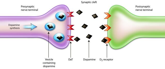
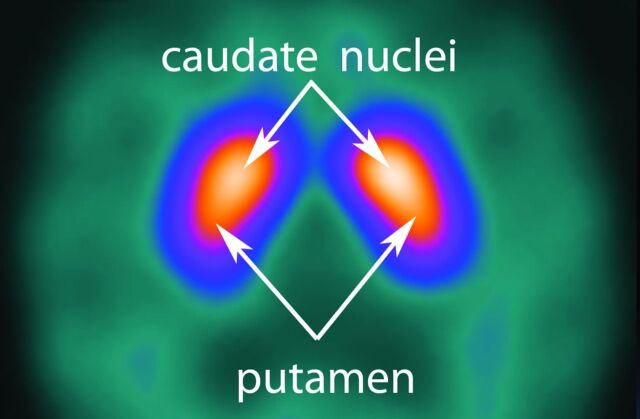
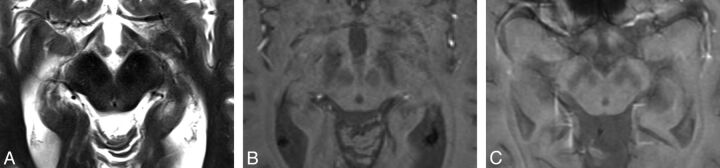
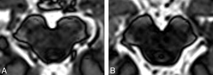
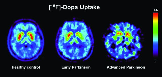
As we defeat infectious diseases and cancer, one of the greatest medical challenges facing us in the mid-21st century will be the increasing prevalence of degenerative disease. Those diseases, which affect movement and cognition, can be the most debilitating. Dysfunction of the extrapyramidal system results in increasing motor disability often manifest as tremor, bradykinesia, and rigidity. The common pathologic pathway of these diseases, collectively described as parkinsonian syndromes, such as Parkinson disease, multiple system atrophy, progressive supranuclear palsy, corticobasal degeneration, and dementia with Lewy bodies, is degeneration of the presynaptic dopaminergic pathways in the basal ganglia. Conventional MR imaging is insensitive, especially in early disease, so functional imaging has become the primary method used to differentiate a true parkinsonian syndrome from vascular parkinsonism, drug-induced changes, or essential tremor. Unusually for a modern functional imaging technique, the method most widely used in European clinics depends on SPECT and not PET. This SPECT technique (described in the first of 2 parts) commonly reports dopamine-transporter function, with decreasing striatal uptake demonstrating increasingly severe disease.


Figures

Fig 1.
Fig 1.
Schematic illustration of the dopaminergic pathway within the striatum. Dopamine is synthesized and stored in vesicles until released into the synaptic cleft in response to an action potential. After interacting with the postsynaptic dopamine receptors, dopamine is released back into the synaptic cleft, where it is actively taken up by dopamine transporters and carried into the presynaptic neuron.
Fig 2.
Fig 2.
Normal study findings. The axial 123I-FP-CIT DaT-SPECT image demonstrates symmetric tracer uptake in the caudate nuclei and putamina, with very low-grade, almost absent, background activity.
Fig 3.
Fig 3.
Axial T2-weighted MR imaging sequence in a patient with PD (A) demonstrating “smudging” of the hypointensity in the substantia nigra toward the red nucleus in the midbrain (right subtlely more marked than the left, concordant with the contralateral clinical features). It is more easily visualized in the corresponding susceptibility-weighted image (B). Axial T2*-weighted MR imaging sequence also demonstrates the “smudging” sign as shown in another patient with PD (C) (left more than right concordant with worse contralateral clinical features). The “smudging” sign is nonspecific and seen in progressive supranuclear palsy, multiple system atrophy, and corticobasal degeneration. It is a subtle sign and often not used in routine clinical practice.
Fig 4.
Fig 4.
On these inversion-recovery T1-weighted images in which deep gray matter signal is suppressed, the substantia nigra in a patient with severe PD (A) appears both substantially shrunk and with altered contrast in comparison with a healthy control (B). There is a correlation between the substantia nigra area with the Unified Parkinson Disease Rating Scale score. There is also a group difference between those with PD and controls; however, this metric has not been proved to be useful for individuals. Images courtesy of Dr Ludovico Minati, Scientific Department, Istituto Di Ricovero e Cura a Carattere Scientifico Foundation Neurologic Institute, Carlo Besta, Milan, Italy.
Fig 5.
Fig 5.
T2-weighted fast spin-echo MR image (A) and corresponding transcranial sonography images (B and C) of midbrain axial sections at the orbitomeatal line. B, Transcranial sonography image of the axial midbrain section in a healthy person depicting the hypoechoic midbrain (outlined) surrounded by the hyperechogenic basal cisterns. In the anatomic area of the ipsilateral substantia nigra, only small hyperechoic patches are visible (small arrows); the planimetrically measured area is <0.20 cm2. The raphe is detected as a highly echogenic, continuous line; the aqueduct is shown at the dorsal part of the midbrain. C, Transcranial sonography of a patient with Parkinson disease showing a bilateral hyperechogenic substantia nigra (area ≥0.20 cm2). Small arrows indicate the substantia nigra. Images courtesy of Dr Rita CL Fernandes, Department of Neurodegeneration at Tübingen University Hospital and Hertie Institute of Clinical Brain Research, Tübingen, Germany.
Fig 6.
Fig 6.
Axial 18F-DOPA PET images through the striatum. The patient with early Parkinson disease shows an asymmetric reduction in putaminal radiotracer uptake. With further disease progression, both putamina show a substantial reduction in radiotracer uptake. Images courtesy of Professor Philippe Remy, l'Hôpital Henri Mondor, Creteil, France.
Fig 7.
Fig 7.
Axial 123I-FP-CIT DaT-SPECT sections depicting the different patterns of abnormality seen in PD as described by Catafau and Tolosa—type 1: asymmetric activity with reduced putaminal uptake in 1 hemisphere (A); type 2: symmetric reduction in putaminal uptake in both hemispheres (B); and type 3: virtual absence of uptake in the putamina and caudate nuclei despite high gain as demonstrated by ample background activity (C).

References

    1. Scherfler C, Schwarz J, Antonini A, et al. Role of DAT-SPECT in the diagnostic work-up of parkinsonism. Mov Disord 2007;22:1229–38 - PubMed
    1. Catafau AM, Tolosa E. Impact of dopamine transporter SPECT using 123I-ioflupane on diagnosis and management of patients with clinically uncertain parkinsonian syndromes. Mov Disord 2004;19:1175–82 - PubMed
    1. Hughes AJ, Daniel SE, Kilford L, et al. Accuracy of clinical diagnosis of idiopathic Parkinson's disease: a clinicopathological study of 100 cases. J Neurol Neurosurg Psychiatry 1992;55:181–84 - PMC - PubMed
    1. Booij J, Tissingh G, Winogrodzka A, et al. Imaging of the dopaminergic neurotransmission system using single-photon emission tomography and positron emission tomography in patients with parkinsonism. Eur J Nucl Med 1999;26:171–82 - PubMed
    1. Seppi K. MRI for the differential diagnosis of neurodegenerative parkinsonism in clinical practice. Parkinsonism Relat Disord 2007;13:S400–05 - PubMed

Publication types

MeSH terms

Substances