. 2015 Jan;26(1):209-19.
doi: 10.1681/ASN.2013121339. Epub 2014 Jun 5.

A novel atypical hemolytic uremic syndrome-associated hybrid CFHR1/CFH gene encoding a fusion protein that antagonizes factor H-dependent complement regulation

Affiliations

A novel atypical hemolytic uremic syndrome-associated hybrid CFHR1/CFH gene encoding a fusion protein that antagonizes factor H-dependent complement regulation

Elisabetta Valoti et al. J Am Soc Nephrol. 2015 Jan.

Abstract

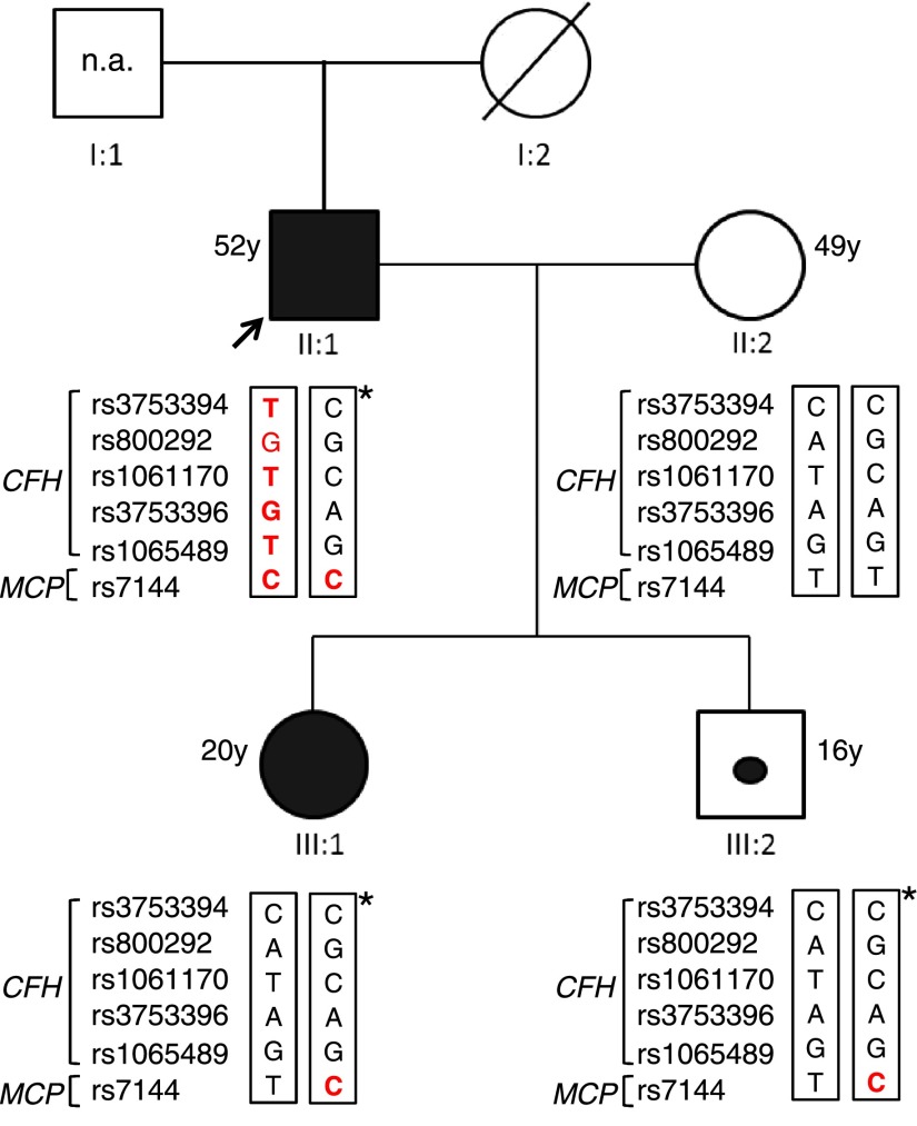
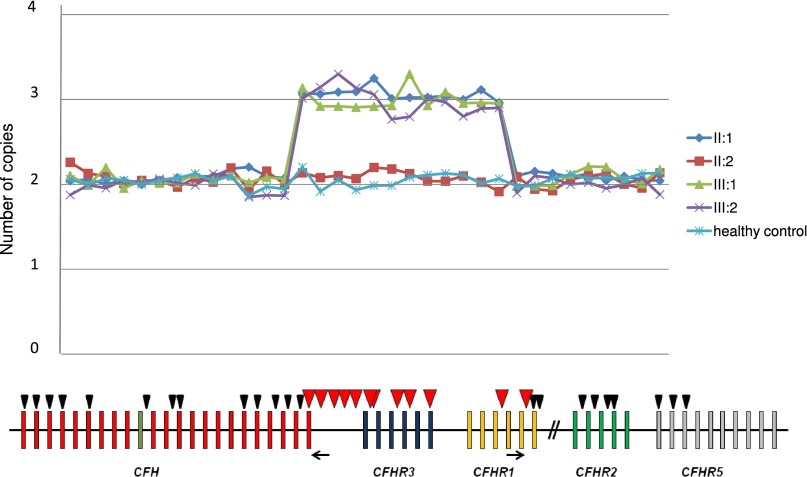
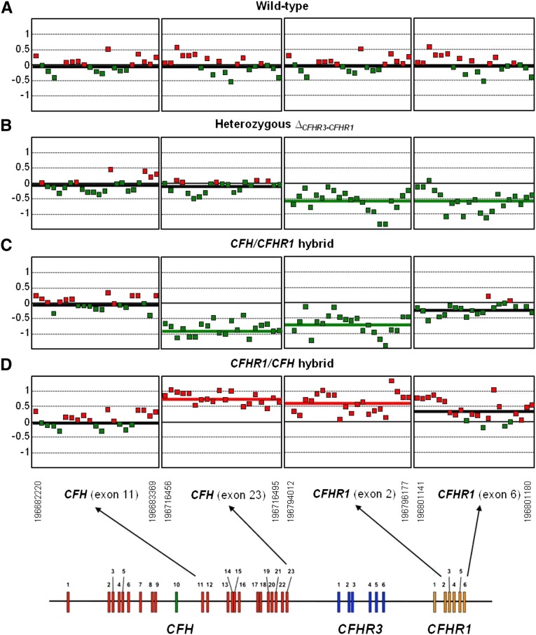
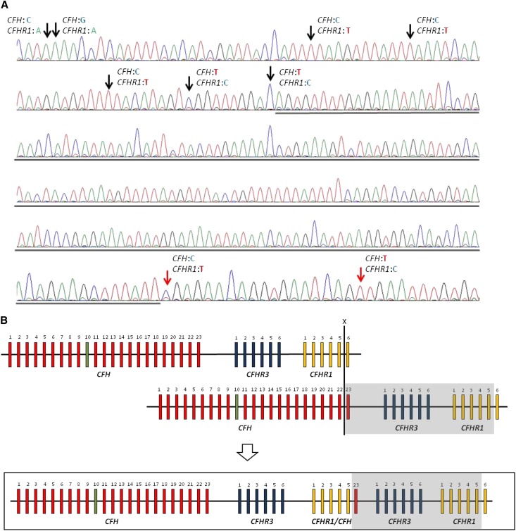
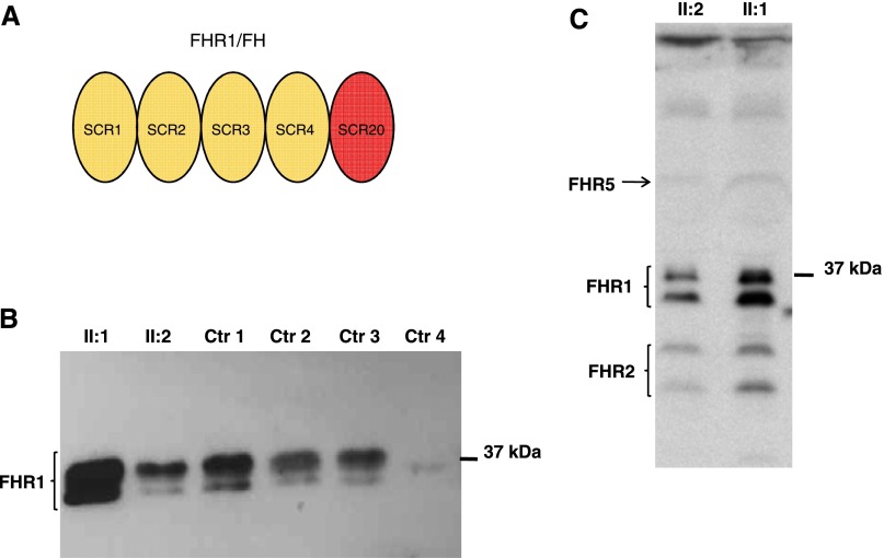
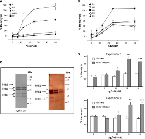
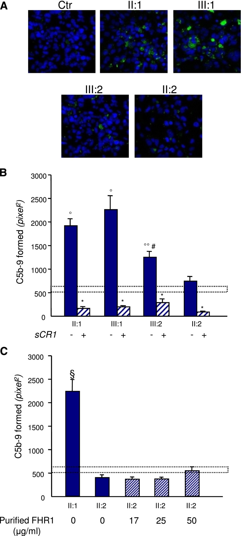
Genomic aberrations affecting the genes encoding factor H (FH) and the five FH-related proteins (FHRs) have been described in patients with atypical hemolytic uremic syndrome (aHUS), a rare condition characterized by microangiopathic hemolytic anemia, thrombocytopenia, and ARF. These genomic rearrangements occur through nonallelic homologous recombinations caused by the presence of repeated homologous sequences in CFH and CFHR1-R5 genes. In this study, we found heterozygous genomic rearrangements among CFH and CFHR genes in 4.5% of patients with aHUS. CFH/CFHR rearrangements were associated with poor clinical prognosis and high risk of post-transplant recurrence. Five patients carried known CFH/CFHR1 genes, but we found a duplication leading to a novel CFHR1/CFH hybrid gene in a family with two affected subjects. The resulting fusion protein contains the first four short consensus repeats of FHR1 and the terminal short consensus repeat 20 of FH. In an FH-dependent hemolysis assay, we showed that the hybrid protein causes sheep erythrocyte lysis. Functional analysis of the FHR1 fraction purified from serum of heterozygous carriers of the CFHR1/CFH hybrid gene indicated that the FHR1/FH hybrid protein acts as a competitive antagonist of FH. Furthermore, sera from carriers of the hybrid CFHR1/CFH gene induced more C5b-9 deposition on endothelial cells than control serum. These results suggest that this novel genomic hybrid mediates disease pathogenesis through dysregulation of complement at the endothelial cell surface. We recommend that genetic screening of aHUS includes analysis of CFH and CFHR rearrangements, particularly before a kidney transplant.

Keywords: complement; genetic renal disease; hemolytic uremic syndrome; kidney disease; transplantation.


Figures

Figure 1.
Figure 1.
Italian family pedigree. The proband (black arrow) is patient II:1, his wife is II:2, his affected daughter is III:1, and the unaffected son is III:2. Patients II:1 and III:1 are represented as a black square and circle, respectively. The unaffected carrier III:2 is indicated by a black dot. Genotype of CFH single nucleotide polymorphisms targeting the CFH-H3 risk (TGTGT) haplotype (rs3753394 c.1–332 C>T, rs800292 c.184G>A p.V62I, rs1061170 c.1204T>C p.Y402H, rs3753396 c.2016A>G p.Q672Q, and rs1065489 c.2808 G>T p.E936D) and the MCP single nucleotide polymorphism (rs7144 c.*897 T>C) targeting the MCPggaac risk haplotype are reported in red. The age of all subjects is shown. n.a., samples not available. *Chromosome with the duplication.
Figure 2.
Figure 2.
Results of MLPA analysis showing a novel CFHR1/CFH hybrid gene. MLPA analysis over the CFH-CFHR region shows three copies in a large region beginning with CFH exon 23 and ending after CFHR1 exon 5 in patients II:1 (proband), III:1 (affected daughter), and III:2 (unaffected son) but not II:2 (unaffected wife). The analysis has been performed with the SALSA MLPA P236-A3 ARMD Kit (MCR Holland) implemented with homemade probes analyzed in a separate assay and covering the last exons and introns of the CFH gene (exon 19, intron 20, intron 21, intron 22, and exon 23). MLPA probes are represented by triangles. Red triangles indicate duplicated probes. The positions of primers for breakpoint mapping are shown by black arrows.
Figure 3.
Figure 3.
CGH array analysis confirms the presence of the CFHR1/CFH hybrid gene. DNAs from four different individuals are compared: (A) a wild-type individual (carrying two copies of CFH, CFHR3, and CFHR1), (B) an individual heterozygous for the CFHR3-CFHR1 deletion, (C) a carrier in heterozygosis of the CFH/CFHR1 hybrid gene, and (D) the proband in this study carrying in heterozygosis the new CFHR1/CFH hybrid gene. As indicated in the diagram at the bottom, only the fragments of the array corresponding to CFH exon 11, CFH exon 23, CFHR1 exon 2, and CFHR1 exon 6 are shown. A thick colored line within each fragment indicates the average DNA gain (red) or loss (green). Notice that, in the carrier of the CFHR1/CFH hybrid gene, CFH exon 23 and CFHR1 exon 2 are overrepresented, confirming the heterozygous duplication in this region. For comparison, the reverse situation occurs in the carrier of the CFH/CFHR1 hybrid gene.
Figure 4.
Figure 4.
Identification of the genomic breakpoint. (A) The breakpoint is located in a region of 193 bp (underlined) between intron 5 of CFHR1 and exon 23 of CFH. The limits of the region are identified by single nucleotide polymorphism differences between CFH and CFHR1 indicated by arrows. Black arrows indicate CFHR1-specific single nucleotide polymorphism variants, and red arrows indicate CFH-specific single nucleotide polymorphism variants. (B) NAHR occurring between CFH and CFHR1 at the positions indicated by the X results in the formation of the CFHR1/CFH hybrid gene consisting of the first five exons of CFHR1, exon 23 of CFH, and an extra copy each of CFHR3 and CFHR1 (boxed area). The duplicated region is highlighted in gray.
Figure 5.
Figure 5.
The FHR1/FH hybrid protein. (A) The protein product of the CFHR1/CFH hybrid gene is a five-SCR protein, where SCR1, -2, -3, and -4 are derived from FHR1 and SCR5 is from FH. (B) Western blot of the FHR1/FH hybrid protein was performed using a polyclonal goat anti-FH antibody against sera from the proband with the heterozygous duplication and the hybrid CFHR1/CFH gene (II:1), his wife (II:2) with two normal copies of CFHR1, a control with two copies of CFHR1 (Ctr 1), two controls with one copy of CFHR1 (Ctr 2 and Ctr 3), and a control with zero copies of CFHR1 (Ctr 4). The presence of the FHR1/FH hybrid protein accounts for the increased band density seen in the proband (II:1). (C) Western blot of the FHR1/FH hybrid protein was performed using a mouse anti–FHR1-FHR2-FHR5 mAb against sera from the proband (II:1) and his wife (II:2). The densities of the two bands of FHR1 isoforms in the proband serum are more pronounced than the densities in his wife’s serum, suggesting the presence of secreted FHR1/FH hybrid protein.
Figure 6.
Figure 6.
Hemolytic and competition assays show that FHR1/FH antagonizes FH-dependent complement regulation on sheep erythrocytes. (A and B) Lysis of sheep erythrocytes by serum from an aHUS patient with the C-terminal mutation W1183L (HUS2) is compared with that obtained with sera from the proband (II:1; with the heterozygous duplication and the hybrid CFHR1/CFH gene), his affected daughter (III:1) and his unaffected son (III:2; both with the heterozygous duplication and the hybrid CFHR1/CFH gene), his wife (II:2; with two normal copies of CFHR1), and a healthy control (Ctr). Lysis is shown as a function of the volume of serum added and is represented as a percentage, setting to 100% either (A) the lysis obtained with 25% serum from the W1183L mutant patient or (B) the lysis obtained with 25% serum from the proband. Data are means±SDs of triplicates. (C, right panel) Coomassie-stained gel of the final purified FHR1 preparations from heterozygous carriers of the CFHR1/CFH hybrid gene (Hybrid) or normal wild-type controls (WT) used in the assays. (C, left panel) Silver-stained gel of the hybrid FHR1/FH-containing fractions eluted from the gel filtration column (containing FHR1, FHR2, and FHR5). In the Coomassie-stained gel, the purified FHR1 preparations from the carriers of the hybrid protein and healthy subjects were normalized to contain identical concentrations of FHR1 protein. Notice that the relative intensities of the FHR1, FHR2, and FHR5 proteins in this gel indicate that there is apparently a smaller amount of FHR2 and FHR5 compared with FHR1 in the carriers of the CFHR1/CFH hybrid gene. This result is consistent with these individuals carrying an extra copy of the CFHR1 gene. The silver-stained gel shows that the preparations are free from contaminants. (D) FHR1 fractions purified from normal controls (white) or heterozygous carriers of the CFHR1/CFH hybrid gene (gray) were added two different control sera (experiments 1 and 2), in which the native FHR1 protein was removed and functional FH was titrated to about 50% by using the anti–N-terminal FH antibody OX24 (6.5 μg/ml OX24 in 10% normal human serum). In both experiments, adding purified FHR1 from heterozygous carriers of the CFHR1/CFH hybrid gene resulted in dose-dependent sheep erythrocyte hemolysis, whereas purified FHR1 from normal serum had no effect. Data are means±SDs of triplicates. *P<0.05, **P<0.01, ***P<0.001 versus WT (purified FHR1 from normal serum).
Figure 7.
Figure 7.
Sera from CFHR1/CFH carriers cause C5b-9 deposition on endothelial cells. Panel A shows representative confocal microscopy images of C5b-9 staining (green) of ADP-activated HMEC-1 exposed for 4 hours to serum (diluted 1:2 in test medium) from a healthy control (Ctr), the proband (II:1; with the heterozygous duplication and the hybrid CFHR1/CFH gene), his affected daughter (III:1) and his unaffected son (III:2; both with the heterozygous duplication and the hybrid CFHR1/CFH gene), and his wife (II:2; with two normal copies of CFHR1). Original magnification, ×400. Blue indicates the 4′,6-diamidino-2-phenylindole staining of cell nuclei. (B) The graph shows the quantification of HMEC-1 area covered by C5b-9 deposits after incubation with serum from a healthy control, the proband, and relatives of the proband in the presence or not of the complement inhibitor sCR1 (150 µg/ml). The dotted rectangle shows the range of C5b-9 deposits induced by a pool of control sera. Data are means±SEMs. *P<0.001 versus serum untreated; °P<0.001, °°P<0.05 versus control; #P<0.05 versus patients. (C) Quantification of HMEC-1 area covered by C5b-9 deposits after incubation with serum from the proband or the healthy wife of the proband with or without the addition of increasing amounts of purified FHR1 from normal serum to mimic the product of one (17 and 25 μg/ml) or two (50 μg/ml) extra CFHR1 gene copies (details are in Supplemental Material). FHR1 addition to serum from the proband’s wife had no effect on C5b-9 deposits. Data are means±SEMs. The dotted rectangle shows the range of C5b-9 deposits induced by a pool of control sera. §P<0.001 versus all the others and versus controls.

References

    1. Noris M, Remuzzi G: Atypical hemolytic-uremic syndrome. N Engl J Med 361: 1676–1687, 2009 - PubMed
    1. Kavanagh D, Goodship TH: Atypical hemolytic uremic syndrome. Curr Opin Hematol 17: 432–438, 2010 - PubMed
    1. Bresin E, Rurali E, Caprioli J, Sanchez-Corral P, Fremeaux-Bacchi V, Rodriguez de Cordoba S, Pinto S, Goodship TH, Alberti M, Ribes D, Valoti E, Remuzzi G, Noris M, European Working Party on Complement Genetics in Renal Diseases : Combined complement gene mutations in atypical hemolytic uremic syndrome influence clinical phenotype. J Am Soc Nephrol 24: 475–486, 2013 - PMC - PubMed
    1. Pérez-Caballero D, González-Rubio C, Gallardo ME, Vera M, López-Trascasa M, Rodríguez de Córdoba S, Sánchez-Corral P: Clustering of missense mutations in the C-terminal region of factor H in atypical hemolytic uremic syndrome. Am J Hum Genet 68: 478–484, 2001 - PMC - PubMed
    1. Dragon-Durey MA, Loirat C, Cloarec S, Macher MA, Blouin J, Nivet H, Weiss L, Fridman WH, Frémeaux-Bacchi V: Anti-Factor H autoantibodies associated with atypical hemolytic uremic syndrome. J Am Soc Nephrol 16: 555–563, 2005 - PubMed

Publication types

Substances

LinkOut - more resources