. 2014 Oct;25(10):2177-86.
doi: 10.1681/ASN.2013070758. Epub 2014 Jun 5.

Blood kidney injury molecule-1 is a biomarker of acute and chronic kidney injury and predicts progression to ESRD in type I diabetes

Affiliations

Blood kidney injury molecule-1 is a biomarker of acute and chronic kidney injury and predicts progression to ESRD in type I diabetes

Venkata S Sabbisetti et al. J Am Soc Nephrol. 2014 Oct.

Abstract

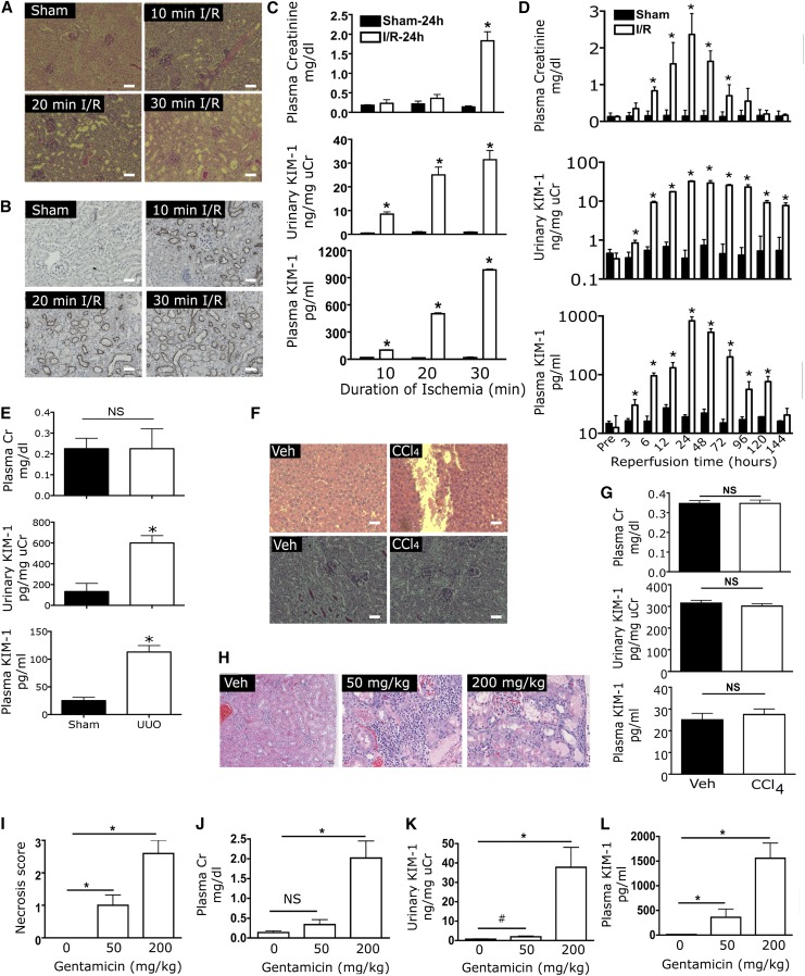
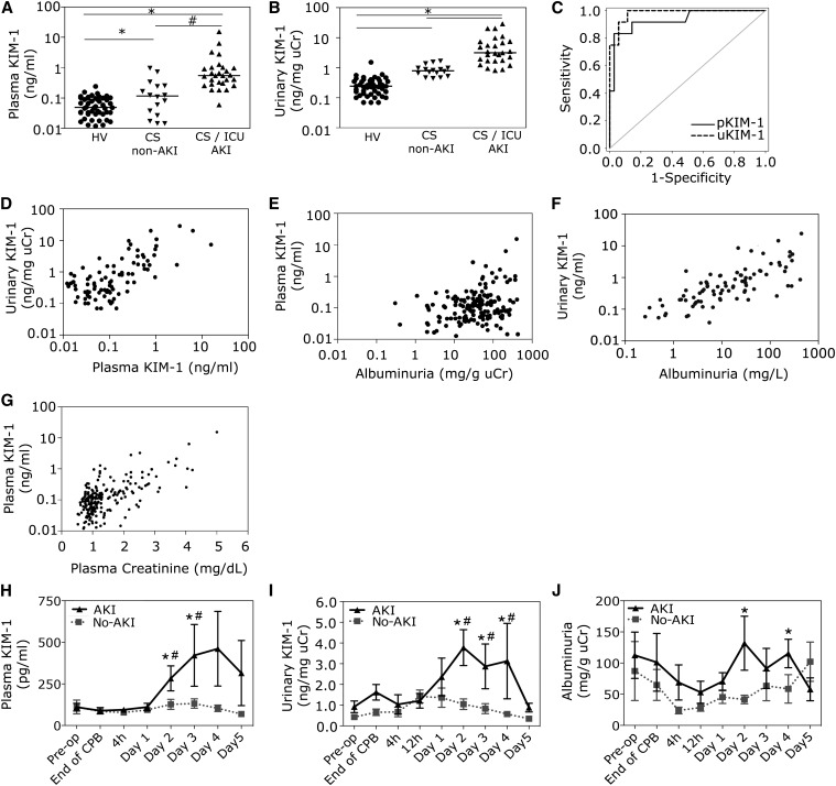
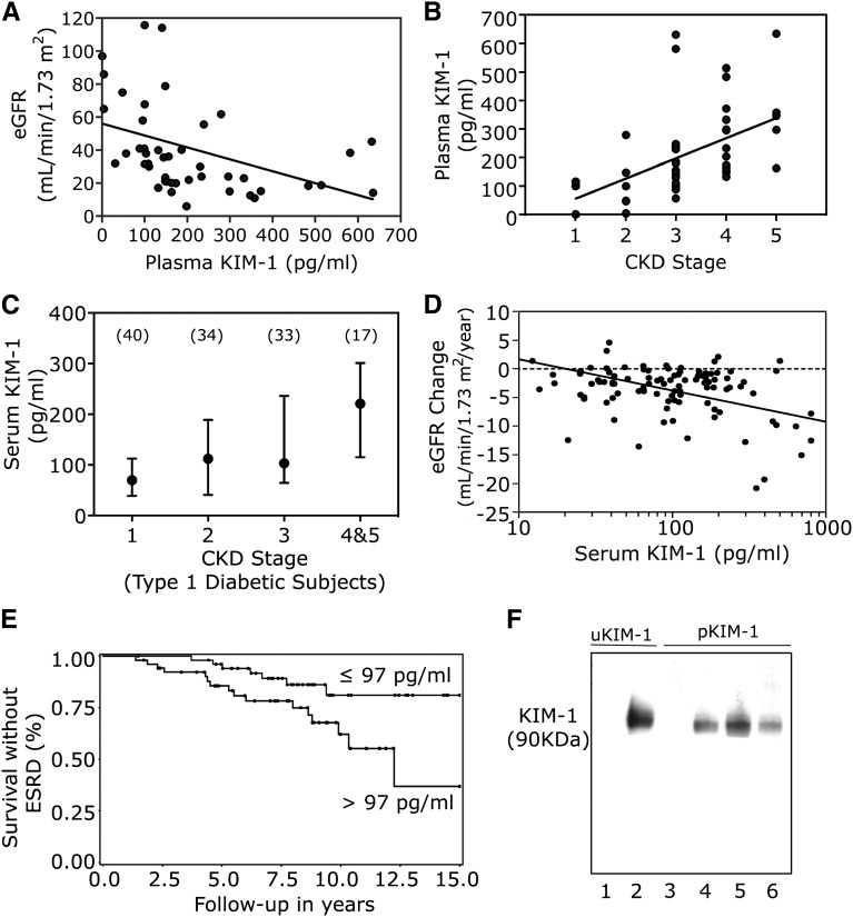
Currently, no blood biomarker that specifically indicates injury to the proximal tubule of the kidney has been identified. Kidney injury molecule-1 (KIM-1) is highly upregulated in proximal tubular cells following kidney injury. The ectodomain of KIM-1 is shed into the lumen, and serves as a urinary biomarker of kidney injury. We report that shed KIM-1 also serves as a blood biomarker of kidney injury. Sensitive assays to measure plasma and serum KIM-1 in mice, rats, and humans were developed and validated in the current study. Plasma KIM-1 levels increased with increasing periods of ischemia (10, 20, or 30 minutes) in mice, as early as 3 hours after reperfusion; after unilateral ureteral obstruction (day 7) in mice; and after gentamicin treatment (50 or 200 mg/kg for 10 days) in rats. In humans, plasma KIM-1 levels were higher in patients with AKI than in healthy controls or post-cardiac surgery patients without AKI (area under the curve, 0.96). In patients undergoing cardiopulmonary bypass, plasma KIM-1 levels increased within 2 days after surgery only in patients who developed AKI (P<0.01). Blood KIM-1 levels were also elevated in patients with CKD of varous etiologies. In a cohort of patients with type 1 diabetes and proteinuria, serum KIM-1 level at baseline strongly predicted rate of eGFR loss and risk of ESRD during 5-15 years of follow-up, after adjustment for baseline urinary albumin-to-creatinine ratio, eGFR, and Hb1Ac. These results identify KIM-1 as a blood biomarker that specifically reflects acute and chronic kidney injury.

Keywords: acute renal failure; chronic kidney disease; chronic kidney failure; nephrotoxicity.


Figures

Figure 1.
Figure 1.
Increase in plasma KIM-1 levels in experimental models of kidney injury in mice and rats. (A) Male BALB/c mice were subjected to 0 (sham), 10, 20, or 30 minutes of bilateral ischemia by clamping the renal pedicles for the time indicated. Urine, blood, and tissue were collected 24 hours after reperfusion. Periodic acid-Schiff staining of kidney sections indicated no injury in sham-operated mice, whereas loss of brush border, necrosis, and sloughing of cells into the tubular lumen were found in postischemic mice. (B) Immunohistochemical staining of KIM-1 on kidney tissues obtained from sham-operated mice and mice that underwent 10, 20, and 30 minutes of ischemia/reperfusion. (C) Plasma creatinine and urinary and plasma KIM-1 in mice 24 hours after challenge with different durations of bilateral ischemia (n=6 per group). (D) Plasma creatinine and urinary and plasma KIM-1 levels were assessed in male BALB/c mice at different times after sham surgery or after reperfusion following 30 minutes of bilateral ischemia (n=6 per group). (E) Male BALB/c mice were subjected to UUO by obstructing the ureter. Urine and blood were collected on day 7 after UUO. Plasma creatinine, urinary KIM-1, and plasma KIM-1 were measured (n=4 per group) *P<0.001. (F) Male BALB/c mice were administered one dose of 10% CCl4 (0.5 ml/kg). Mice were euthanized 48 hours after CCl4 administration and evaluated for liver (upper two panels) or kidney (lower two panels) toxicity by histopathology after periodic acid-Schiff staining (n=3 per group). (G) Plasma creatinine, normalized urinary KIM-1, or plasma KIM-1 concentration in vehicle (Veh) and CCl4-treated mice (n=3 per group) *P<0.001. (H) Male Sprague-Dawley rats were administrated 0.9% saline or 50 or 200 mg/kg gentamicin daily for 10 days and euthanized on day 11. Hematoxylin and eosin staining of kidney sections revealed no injury in vehicle-treated rats, whereas there were loss of brush border, necrosis, and sloughing of cells into the tubular lumen in gentamicin-treated rats. Tubular necrosis score (I), plasma creatinine (J), urinary KIM-1 normalized to urine creatinine (K), and plasma KIM-1 (L) in rats administrated gentamicin at 0, 50, or 200 mg/kg per day for 10 days. *P<0.001, #P<0.05; n=5). Scale bars, 50 μm. Error bars reflect SEM.
Figure 2.
Figure 2.
Plasma KIM-1 is a marker of renal injury in human AKI. Plasma and urine were collected from healthy volunteers and post–cardiac surgery (CS) patients with or without AKI and ICU patients with AKI from other causes. Dot plots indicate plasma KIM-1 (A) and urinary KIM-1 normalized to urinary creatinine (B) for each patient. *P<0.001; #P<0.05. (C) ROC curve analysis comparing performance of normalized urinary KIM-1 (dashed red line, AUC 0.98) and plasma KIM-1 (solid black line, AUC 0.96) levels. (D) Scatter plot demonstrating a positive correlation between plasma and urinary KIM-1 levels in all participants, including healthy volunteers (n=48) and patients with (n=28) or without AKI (n=16). (r=0.43; P<0.001). (E) Scatterplot demonstrating a correlation between plasma KIM-1 levels and urinary albumin-to-creatinine ratios (r=0.33; P=0.001). (F) Scatter plot demonstrating a correlation between urinary KIM-1 levels and urinary albumin levels (r=0.35; P<0.001 for urinary KIM-1). (G) Scatter plot demonstrating positive correlation between plasma KIM-1 and plasma creatinine in patients with or without AKI (r=0.58; P<0.001). (H) Plasma and urine were collected at various times before and after CPB from nine patients who developed stage 1 AKI and nine who did not develop AKI. Mean plasma creatinine, plasma KIM-1 (H), normalized urinary KIM-1 (I), and urinary albumin (J) concentrations were determined. #P<0.05 for difference from baseline; *P<0.05 for difference between AKI and non-AKI groups. n=9 for both AKI and no-AKI groups. Error bars represents SDs.
Figure 3.
Figure 3.
Blood KIM-1 as a biomarker of CKD and predictor of progression of patients with type 1 diabetes. (A) In a cross-sectional comparison, plasma KIM-1 levels were negatively associated with eGFR in patients with CKD of various causes. (B) Plasma KIM-1 levels are positively associated with increasing stages of CKD. (C) In a cross-sectional comparison of 124 patients with type 1 diabetes and proteinuria, serum KIM-1 was positively associated with CKD stage. Median and 25th and 75th percentiles are shown. Numbers of patients in each category are indicated. (D) Serum KIM-1 at baseline was associated with rate of renal decline (eGFR slopes) during 5–15 years (median, 10 years) of follow-up (Spearman correlation coefficient=0.52; P<0.001). The effect of serum KIM-1 remained very strong and significant (P<0.001) in multiple regression analyses when other covariates, such as baseline eGFR, urinary albumin-to-creatinine ratio, and hemoglobin A1c levels were considered. (E) Serum KIM-1 level at baseline was a strong predictor of risk of progression to ESRD. Kaplan–Meier survival analysis shows the proportion of patients remaining without ESRD after 15 years of follow-up in patients with baseline serum KIM-1 below and above the median (97 pg/ml) (P<0.01). The effect of baseline serum KIM-1 remained significant in multivariable Cox regression analysis (P<0.01) when other covariates, such as baseline eGFR, urinary albumin-to-creatinine ratio, and hemoglobin A1c levels were considered. Analyses shown in D and E were performed in 107 patients with type 1 diabetes, proteinuria, and CKD stages 1–3 at baseline. More clinical information of these patients is provided in Supplemental Table 4 and can be found in Rosolowsky et al. (F) Western blot depicting 90-kD band of urinary KIM-1 in a patient with AKI (lane 2) and plasma KIM-1 in patients with AKI (lanes 4 and 5), and CKD (lane 6). Urine (lane 1) and plasma (lane 3) from healthy volunteers were also included for comparision.

References

    1. Ichimura T, Bonventre JV, Bailly V, Wei H, Hession CA, Cate RL, Sanicola M: Kidney injury molecule-1 (KIM-1), a putative epithelial cell adhesion molecule containing a novel immunoglobulin domain, is up-regulated in renal cells after injury. J Biol Chem 273: 4135–4142, 1998 - PubMed
    1. Han WK, Bailly V, Abichandani R, Thadhani R, Bonventre JV: Kidney Injury Molecule-1 (KIM-1): a novel biomarker for human renal proximal tubule injury. Kidney Int 62: 237–244, 2002 - PubMed
    1. Ichimura T, Hung CC, Yang SA, Stevens JL, Bonventre JV: Kidney injury molecule-1: a tissue and urinary biomarker for nephrotoxicant-induced renal injury. Am J Physiol Renal Physiol 286: F552–F563, 2004 - PubMed
    1. Vaidya VS, Ozer JS, Dieterle F, Collings FB, Ramirez V, Troth S, Muniappa N, Thudium D, Gerhold D, Holder DJ, Bobadilla NA, Marrer E, Perentes E, Cordier A, Vonderscher J, Maurer G, Goering PL, Sistare FD, Bonventre JV: Kidney injury molecule-1 outperforms traditional biomarkers of kidney injury in preclinical biomarker qualification studies. Nat Biotechnol 28: 478–485, 2010 - PMC - PubMed
    1. Vaidya VS, Waikar SS, Ferguson MA, Collings FB, Sunderland K, Gioules C, Bradwin G, Matsouaka R, Betensky RA, Curhan GC, Bonventre JV: Urinary biomarkers for sensitive and specific detection of acute kidney injury in humans. Clin Transl Sci 1: 200–208, 2008 - PMC - PubMed

Publication types

MeSH terms