. 2015 Jan;26(1):67-80.
doi: 10.1681/ASN.2013101045. Epub 2014 Jun 5.

Recombination signal binding protein for Ig-κJ region regulates juxtaglomerular cell phenotype by activating the myo-endocrine program and suppressing ectopic gene expression

Affiliations

Recombination signal binding protein for Ig-κJ region regulates juxtaglomerular cell phenotype by activating the myo-endocrine program and suppressing ectopic gene expression

Ruth M Castellanos-Rivera et al. J Am Soc Nephrol. 2015 Jan.

Abstract

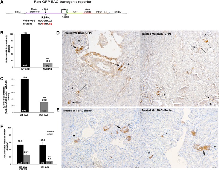
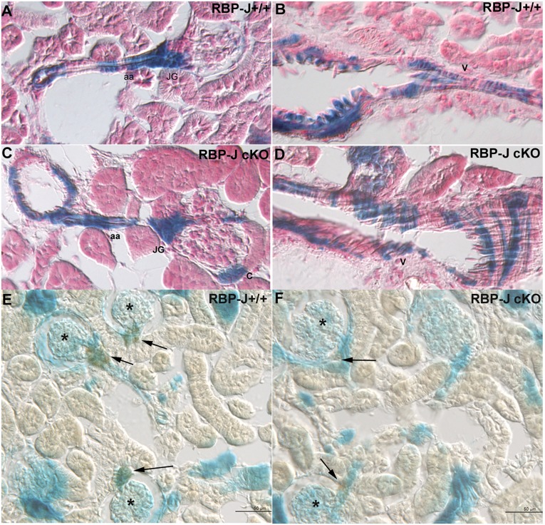
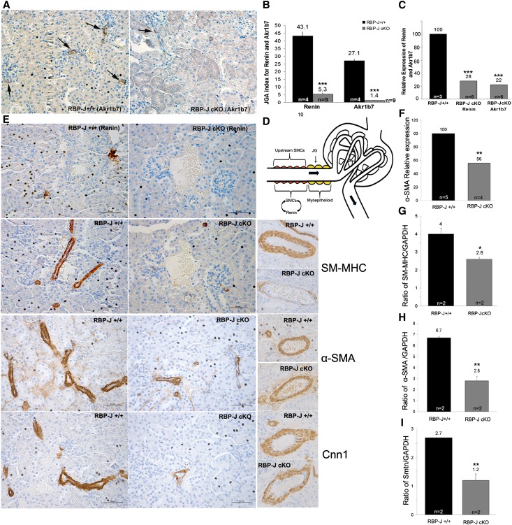
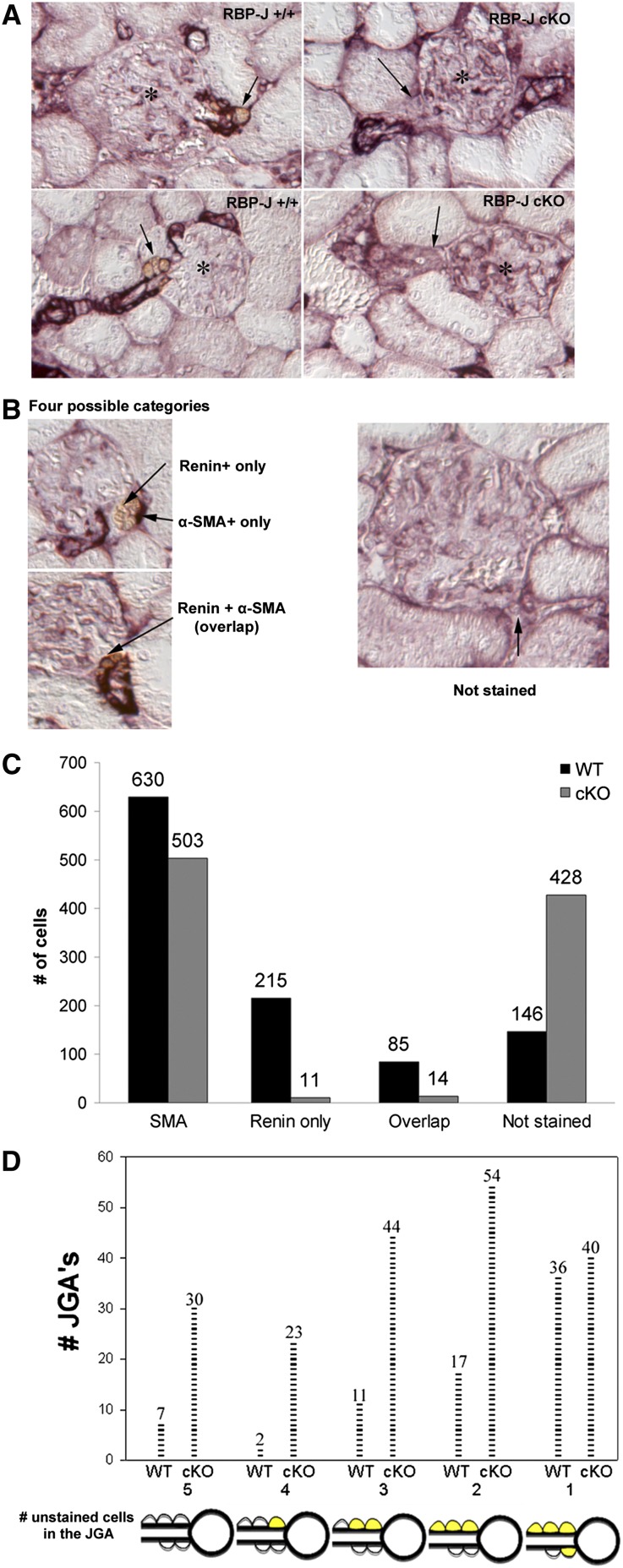
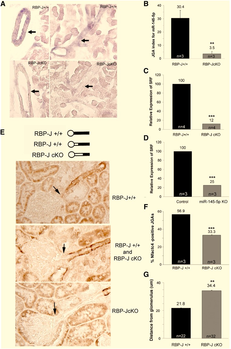
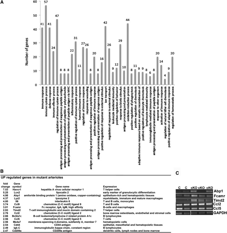
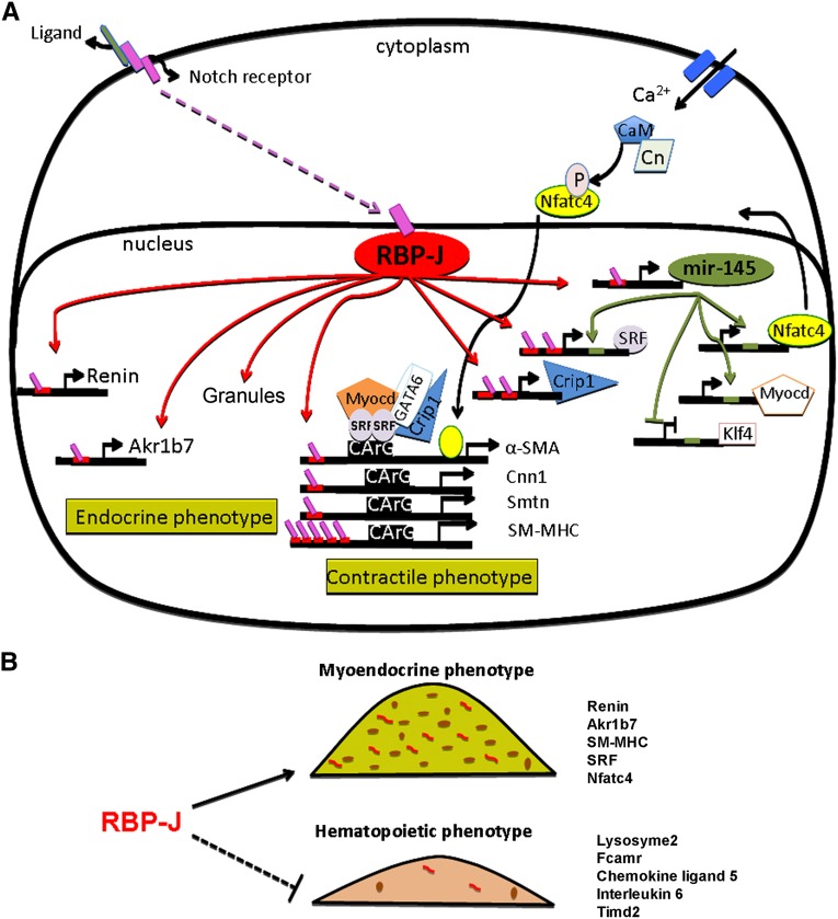
Recombination signal binding protein for Ig-κJ region (RBP-J), the major downstream effector of Notch signaling, is necessary to maintain the number of renin-positive juxtaglomerular cells and the plasticity of arteriolar smooth muscle cells to re-express renin when homeostasis is threatened. We hypothesized that RBP-J controls a repertoire of genes that defines the phenotype of the renin cell. Mice bearing a bacterial artificial chromosome reporter with a mutated RBP-J binding site in the renin promoter had markedly reduced reporter expression at the basal state and in response to a homeostatic challenge. Mice with conditional deletion of RBP-J in renin cells had decreased expression of endocrine (renin and Akr1b7) and smooth muscle (Acta2, Myh11, Cnn1, and Smtn) genes and regulators of smooth muscle expression (miR-145, SRF, Nfatc4, and Crip1). To determine whether RBP-J deletion decreased the endowment of renin cells, we traced the fate of these cells in RBP-J conditional deletion mice. Notably, the lineage staining patterns in mutant and control kidneys were identical, although mutant kidneys had fewer or no renin-expressing cells in the juxtaglomerular apparatus. Microarray analysis of mutant arterioles revealed upregulation of genes usually expressed in hematopoietic cells. Thus, these results suggest that RBP-J maintains the identity of the renin cell by not only activating genes characteristic of the myo-endocrine phenotype but also, preventing ectopic gene expression and adoption of an aberrant phenotype, which could have severe consequences for the control of homeostasis.

Keywords: cell fate; hematopoiesis; juxtaglomerular cell; renin; smooth muscle.


Figures

Figure 1.
Figure 1.
RBP-J regulates the renin promoter in vivo. Mutation of the RBP-J site in the renin promoter diminishes GFP expression, a surrogate of renin expression. (A) Diagram of the BAC reporter construct used to generate BAC transgenic mice. The dual kanamycin/streptomycin selection strategy was used to replace the first exon of renin with enhanced GFP, insert the renin gene 3′ untranslated region (UTR), and modify the RBP-J binding site (nucleotides are in red). (B) Quantitative RT-PCR for GFP mRNA expression in kidneys from mice harboring the mutated RBP-J reporter (Mut-BAC) is significantly lower than in the control (WT-BAC) mice at basal state. (C–F) Mice were treated with low sodium diet+captopril for 5 days to induce re-expression of renin along arterioles. (C) Mut-BAC mice do not increase GFP mRNA to the same level as WT-BAC mice. (D and E) Immunohistochemistry for GFP and renin expression in response to homeostatic challenge. (D) In WT-BAC mice, GFP (brown) is in JG cells, small arterioles, and along the arterioles, recapitulating the renin pattern. Mut-BAC mice have no GFP along the arterioles and few GFP-positive JGAs (arrows). (E) Renin staining in WT-BAC and Mut-BAC mice shows that, as expected, the endogenous, nonmutated renin gene responds by increasing the number of renin-positive cells along the arterioles properly and equally in both groups. *Glomeruli with GFP-positive or renin-positive JGA. (F) The JGA index for renin in both transgenic mice shows that harboring the BAC transgene does not affect expression of the endogenous renin gene. Values are means±SEMs. **P<0.01; ***P<0.001.
Figure 2.
Figure 2.
RBP-J deletion does not affect the endowment of cells from the renin lineage. Kidneys from control and RBP-J cKO;R26R adult mice were subjected to the X-gal reaction to detect β-gal, which marks cells that expressed renin earlier in development (blue). (A and B) In RBP-J+/+ control kidneys, blue staining is seen in the JGA (JG), along the afferent arteriole (aa), and in a large vessel (V). (C and D) RBP-J cKO kidneys have the same blue staining pattern as controls. (E and F) Double staining for β-gal (blue) and renin (brown). Whereas β-gal labels the developmental history of renin expression (thus, blue staining is observed along arterioles and glomeruli), the actual expression of renin (brown) in adult animals is confined to JG cells. In cKO mice, the distribution of blue cells in the JGA and arterioles is not different from controls; however, cKO mice have fewer renin+ JGAs (arrows) than in control mice. *Glomeruli with renin+ JGAs.
Figure 3.
Figure 3.
Deletion of RBP-J affects expression of genes marking the dual endocrine and SM phenotype of renin cells. (A–C) Expression of Akr1b7, an endocrine phenotype marker. (A) Immunostaining for Akr1b7 shows that RBP-J+/+ mice express Akr1b7 in the JGAs (arrows in left panel). RBP-J cKO mice have few or no Akr1b7-positive JGAs (arrow in right panel). (B) In RBP-J cKO mice, the JGA indices for both Akr1b7 and renin are significantly reduced. (C) Akr1b7 and renin mRNA levels in RBP-J cKO mice decreased significantly to the same extent compared with controls. (D–I) Expression of SM phenotype markers assessed by (D and E) immunostaining and (F–I) mRNA expression. (D) The sketch illustrates the location of JG cells (yellow) in the adult animal and the location of VSMCs upstream in the afferent arterioles, where expression of SM genes is reduced in RBP-J cKO mice. The diagram also indicates that the SM cells (SMCs) have the plasticity to re-express the renin phenotype when confronted with a homeostatic threat. (E) Consecutive sections stained with renin and SM-MHC show that RBP-J cKO mice had few renin-positive cells, and the expression of SM-MHC was markedly reduced, particularly in the interlobular arteries and upstream portion of the afferent arterioles. Consecutive sections stained for α-SMA and Cnn1 show that α-SMA staining upstream in the afferent arterioles is thinner with decreased intensity and extent of expression. Cnn1 staining shows an overall decrease in the number of Cnn1-positive cells. In large arteries (right panels), cKO mice had fewer SM-MHC–positive cells around the artery, but localization of α-SMA and Cnn1 was not different from controls. Notice that SM-MHC in mutant mice had a scant and dot-like distribution of SM-MHC–positive cells around the artery. (F) α-SMA mRNA expression was reduced to 56% of control in kidneys from cKO mice. (G–I) Semiquantitative PCR in isolated kidney arterioles showed that α-SMA, SM-MHC, and smoothelin expression decreased 2.4-, 1.5-, and 2.3-fold, respectively, in the RBP-J cKO mice. Values are means±SEMs. Scale bars, 50 µm. *P<0.05; **P<0.01; ***P<0.001.
Figure 4.
Figure 4.
Deletion of RBP-J increases the number of JG cells that express neither renin nor SM markers. (A) Double staining for renin (brown) and α-SMA (purple) shows that cKO mice have few or no renin-positive JG cells (arrows) and diminished or absent α-SMA expression in JGAs. *Glomeruli. (B) Five cells in each JGA were counted and categorized as (1) renin+ only, (2) α-SMA+ only, (3) both renin+ and α-SMA+, and (4) not stained. (C) A summary of the number of cells in each category shows that RBP-J cKO mice had an increased number of unstained cells. (D) The number of JGAs with one to five cells unstained with either antibody. RBP-J cKO mice had more JGAs with two to five unstained cells, whereas the number of JGAs with one unstained cell is similar in RBP-J cKO and control animals. In total, 219 JGA cells were counted in the controls (n=2), and 216 JGA cells were counted in the mutants (n=2). Each tick indicates a JGA. Cells in yellow are cells stained with either antibody.
Figure 5.
Figure 5.
Deletion of RBP-J reduces expression of miR-145–5p and its downstream SM gene targets. (A) In situ hybridization for miR-145–5p. In control mice, miR-145–5p is expressed in SM cells along the arterioles and in major arteries (arrows), whereas the RBP-J cKO mice exhibit a significant decrease in the overall expression of miR-145–5p. (B) In addition, the number of JGAs positive for miR-145–5p was decreased. (C) SRF mRNA expression is reduced significantly in both RBP-J cKO and miR-145–5p KO mice. (D–G) Nfatc4 expression in RBP-J cKO mice. (E) Staining for Nfatc4 showed that some JGAs (arrows) were Nfatc4-positive and that some were negative; however, the upstream portion of the arterioles was positive. The diagram illustrates the distribution of Nfatc4-positive cells. (F) JGAs with a visible arteriole were examined in control and cKO mice, and it was seen that the percent of Nfatc4-positive JGAs is reduced in RBP-J cKO mice. (G) To define whether RBP-J deletion altered the distribution (distance) of Nfatc4-positive cells from the glomerulus, we measured the distance from the JGA to the first Nfatc4-positive cells along the arteriole upstream from the glomerulus. In RBP-J cKO mice, the majority of the Nfatc4-positive cells were farther upstream of the JGA compared with controls. Values are means±SEMs. **P<0.01; ***P<0.001.
Figure 6.
Figure 6.
Genes associated with the hematopoietic immune response are upregulated in arterioles from RBP-J cKO mice. (A) Gene Ontology analysis of the mRNA microarray data in isolated arterioles from RBP-J cKO mice shows increased representation of biologic processes related to the immune response. (B) Immune response-related genes upregulated in RBP-J cKO arterioles. (C) Validation of upregulated genes by PCR of RNA from control and RBP-J cKO kidneys. GAPDH was used for normalization.
Figure 7.
Figure 7.
RBP-J acts as a master regulator that maintains the identity of the JG cell. (A) RBP-J regulates a network of genes that confers the endocrine–contractile phenotype of the JG cell. Red bars located in the promoters of target genes represent RBP-J binding sites. Endocrine genes, such as renin and Akr1b7, and generation of renin granules are indicated on the left side of the diagram. Genes necessary to maintain the contractile phenotype are indicated on the right side. RBP-J regulates the SM genes directly through their promoter regions and indirectly by upregulating the expression of miR-145–5p and SRF. miR-145–5p positively regulates SRF, myocardin, and Nfatc4, and they act together to activate the expression of SM genes. SRF binds to CArG sites located in the SM genes. Given that SRF is an miR-145–5p target gene and has two RBP-J sites in its promoter, it is likely that both miR-145–5p and RBP-J regulate the transcriptional activity of SRF. In addition, miR-145–5p also promotes the expression of SM genes by repressing Klf4, a transcription factor that forms a complex with SRF to prevent transcriptional activity of SM genes. Crip1, another RBP-J predicted target gene, is an SM marker known to form a complex with GATA 6 and SRF to promote the contractile phenotype. We hypothesize that the canonical Notch signaling pathway is involved in maintaining the myo-endocrine phenotype of the JG cell. The ligand–Notch receptor interaction (yet to be identified) results in the release of the Notch intracellular domain (pink boxes), allowing its translocation to the nucleus, where it binds RBP-J to activate transcription. CaM, calmodulin; Cn, calcineurin; P, phosphate; Smtn, smoothelin. (B) RBP-J maintains the identity of the JG cells by not only activating genes characteristic of their myo-endocrine phenotype but also, preventing the undesirable ectopic expression of genes from other lineages.

References

    1. Berg AC, Chernavvsky-Sequeira C, Lindsey J, Gomez RA, Sequeira-Lopez ML: Pericytes synthesize renin. World J Nephrol 2: 11–16, 2013 - PMC - PubMed
    1. Gomez RA, Chevalier RL, Sturgill BC, Johns DW, Peach MJ, Carey RM: Maturation of the intrarenal renin distribution in Wistar-Kyoto rats. J Hypertens 4: S31–S33, 1986
    1. Gomez RA, Lynch KR, Chevalier RL, Wilfong N, Everett A, Carey RM, Peach MJ: Renin and angiotensinogen gene expression in maturing rat kidney. Am J Physiol 254: F582–F587, 1988 - PubMed
    1. Gomez RA, Chevalier RL, Everett AD, Elwood JP, Peach MJ, Lynch KR, Carey RM: Recruitment of renin gene-expressing cells in adult rat kidneys. Am J Physiol 259: F660–F665, 1990 - PubMed
    1. Sequeira Lopez ML, Pentz ES, Robert B, Abrahamson DR, Gomez RA: Embryonic origin and lineage of juxtaglomerular cells. Am J Physiol Renal Physiol 281: F345–F356, 2001 - PubMed

Publication types

MeSH terms

Substances