. 2015 Jan;26(1):48-54.
doi: 10.1681/ASN.2014030265. Epub 2014 Jun 5.

Renin lineage cells repopulate the glomerular mesangium after injury

Affiliations

Renin lineage cells repopulate the glomerular mesangium after injury

Charlotte Starke et al. J Am Soc Nephrol. 2015 Jan.

Abstract

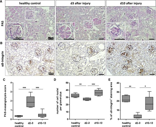
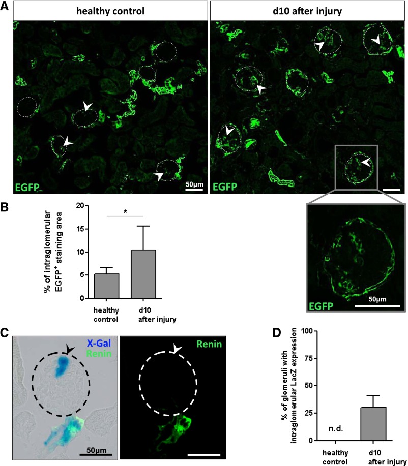
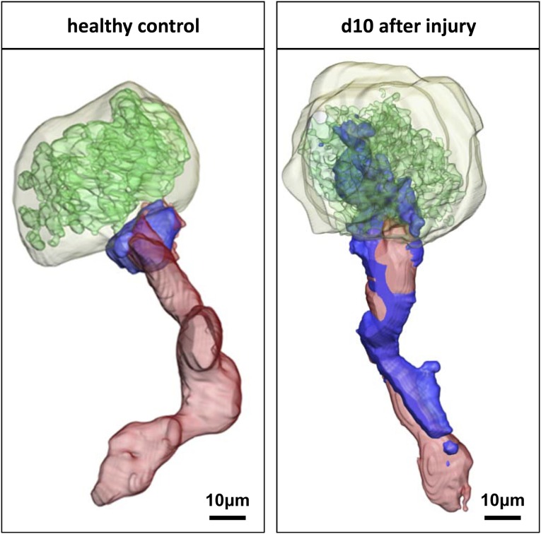
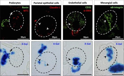
Mesangial cell injury has a major role in many CKDs. Because renin-positive precursor cells give rise to mesangial cells during nephrogenesis, this study tested the hypothesis that the same phenomenon contributes to glomerular regeneration after murine experimental mesangial injury. Mesangiolysis was induced by administration of an anti-mesangial cell serum in combination with LPS. In enhanced green fluorescent protein-reporter mice with constitutively labeled renin lineage cells, the size of the enhanced green fluorescent protein-positive area in the glomerular tufts increased after mesangial injury. Furthermore, we generated a novel Tet-on inducible triple-transgenic LacZ reporter line that allowed selective labeling of renin cells along renal afferent arterioles of adult mice. Although no intraglomerular LacZ expression was detected in healthy mice, about two-thirds of the glomerular tufts became LacZ positive during the regenerative phase after severe mesangial injury. Intraglomerular renin descendant LacZ-expressing cells colocalized with mesangial cell markers α8-integrin and PDGF receptor-β but not with endothelial, podocyte, or parietal epithelial cell markers. In contrast with LacZ-positive cells in the afferent arterioles, LacZ-positive cells in the glomerular tuft did not express renin. These data demonstrate that extraglomerular renin lineage cells represent a major source of repopulating cells for reconstitution of the intraglomerular mesangium after injury.

Keywords: glomerular disease; mesangial cells; stem cell.


Figures

Figure 1.
Figure 1.
Severe mesangial cell injury induced by administration of LPS and anti-mesangial cell serum. (A) Histologic evaluation of PAS staining shows mesangiolysis and decreased glomerular cell number (D) on day 3 after model induction compared with the healthy control. (C) The PAS-mesangiolysis score further indicates mesangial cell loss on days 2–3 followed by hypercellularity on days 10–13 (D). (B and E) Expression of the mesangial cell marker α8-integrin is detected in all glomeruli of healthy control mice. Staining of α8-integrin is significantly reduced on days 2–3 after disease induction but mesangiolysis is not homogenously distributed in all glomeruli on days 2–3. On day 10 after injury, α8-integrin–positive mesangial cells repopulate the glomerular tuft. Box plots show the median with 25th and 75th percentiles and error bars show 10th and 90th percentiles (n=5–8 per group). *P<0.05; **P<0.01; ***P<0.001.
Figure 2.
Figure 2.
Fate-mapped cells of renin lineage are located in the glomerular tuft after experimental mesangial injury. (A) Mesangial injury is induced in mRenCre/tdTomato-EGFP mice to trace renin lineage cells using EGFP as the reporter. No antibody is required to visualize EGFP reporter cells, which localize to the afferent arterioles or parietal cells of Bowman’s capsule. Representative images demonstrate that only a small glomerular area shows EGFP-positive reporter cells within the glomerular tuft in healthy control mice, as indicated by arrowheads (left). On day 10 after disease induction, an increased intraglomerular area displays EGFP-positive reporter cells (right, arrowheads). A higher magnification view of the boxed region shows EGFP-tagged cells with typical localization for mesangial cells on day 10. (B) Statistical analysis of EGFP-positive staining area per glomerulus shows a significantly increased percentage on day 10 after mesangial cell injury compared with healthy controls (n=6–10 per group). (C) To determine whether renin lineage cells are recruited from an extraglomerular origin, experimental mesangial damage is induced in mRen-rtTAm2/LC1/R26R-LacZ mice. Genetically tagged cells of renin lineage are visualized using β-gal staining. A representative glomerulus on day 10 after disease induction shows a considerable fraction of LacZ-expressing cells in the glomerular tuft (left, arrowhead). In contrast with the juxtaglomerular apparatus, intraglomerular β-gal–stained cells do not overlap with renin staining (right). (D) The percentage of glomeruli with intraglomerular LacZ-expressing cells per cross-section is markedly increased in mice on day 10 after injury (n=7) compared with healthy controls (n=5). Data represent the mean±SD. *P<0.05. n.d., not detectable.
Figure 3.
Figure 3.
Extraglomerular origin of repopulating renin lineage cells after mesangial injury. Three-dimensional glomerular reconstruction of α-smooth muscle actin (red, afferent arterioles), α8-integrin–immunoreactive (green), and LacZ (blue) positive areas in healthy controls (left, no intraglomerular staining) of mRen-rtTAm2/LC1/R26RLacZ mice and on day 10 (right, positive intraglomerular staining) after experimental mesangial injury. All animals receive doxycycline and are treated with enalapril.
Figure 4.
Figure 4.
Cells of renin lineage are precursors of regenerating mesangial cells. Immunohistochemical and β-gal costaining is performed to determine whether recruited intraglomerular cells of renin lineage express proteins that are considered to be podocyte-, parietal epithelial-, endothelial-, or mesangial cell-specific markers. Representative images of intraglomerular LacZ-expressing cells (blue, arrowheads) on day 10 after mesangial disease induction show no coexpression with WT1 (red, podocytes), renin (green), Claudin-1 (red, parietal epithelial cells), CD31 (green, endothelial cells), and α-smooth muscle actin (red, α-SMA). Costaining of the mesangial cell marker α8-integrin and β-gal reveals colocalization of the genetically labeled renin lineage cells in the glomerular tuft.

References

    1. Schlöndorff D, Banas B: The mesangial cell revisited: No cell is an island. J Am Soc Nephrol 20: 1179–1187, 2009 - PubMed
    1. Morita T, Churg J: Mesangiolysis. Kidney Int 24: 1–9, 1983 - PubMed
    1. Barratt J, Feehally J: IgA nephropathy. J Am Soc Nephrol 16: 2088–2097, 2005 - PubMed
    1. Hagemann JH, Haegele H, Müller S, Anders HJ: Danger control programs cause tissue injury and remodeling. Int J Mol Sci 14: 11319–11346, 2013 - PMC - PubMed
    1. Hugo C, Shankland SJ, Bowen-Pope DF, Couser WG, Johnson RJ: Extraglomerular origin of the mesangial cell after injury. A new role of the juxtaglomerular apparatus. J Clin Invest 100: 786–794, 1997 - PMC - PubMed

Publication types

MeSH terms

LinkOut - more resources