. 2014 Jun 6;344(6188):1131-5.
doi: 10.1126/science.1252043.

Structural basis for a pH-sensitive calcium leak across membranes

Affiliations

Structural basis for a pH-sensitive calcium leak across membranes

Yanqi Chang et al. Science. .

Abstract

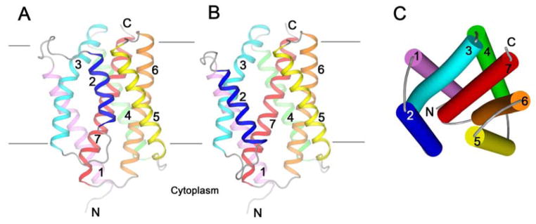
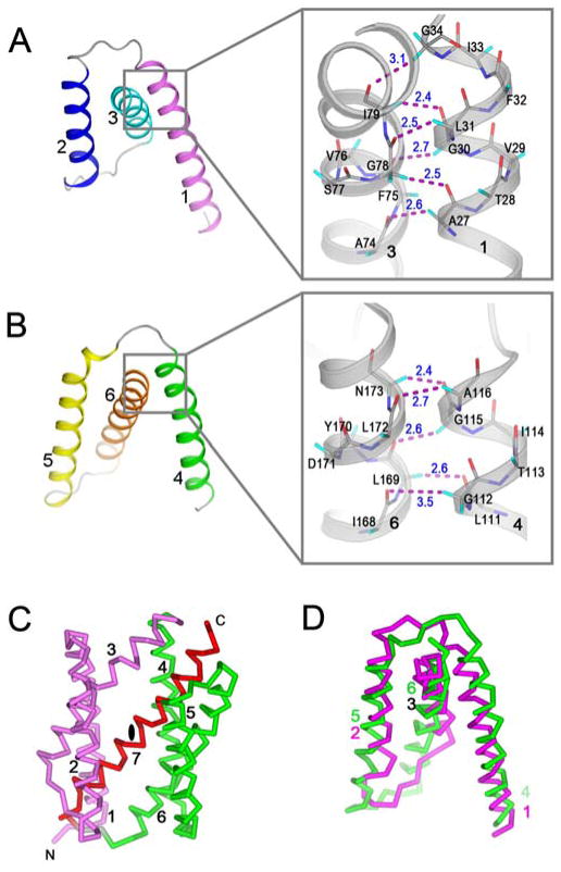
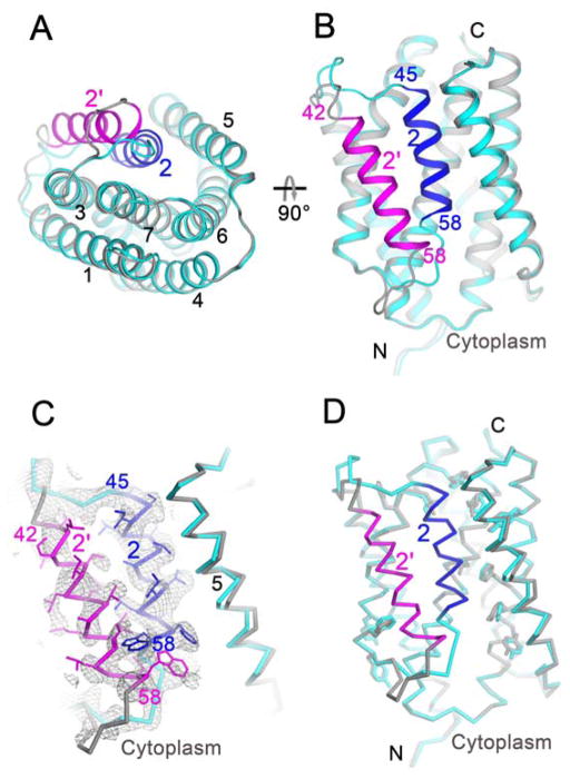
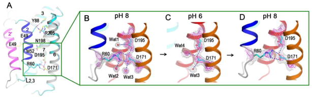
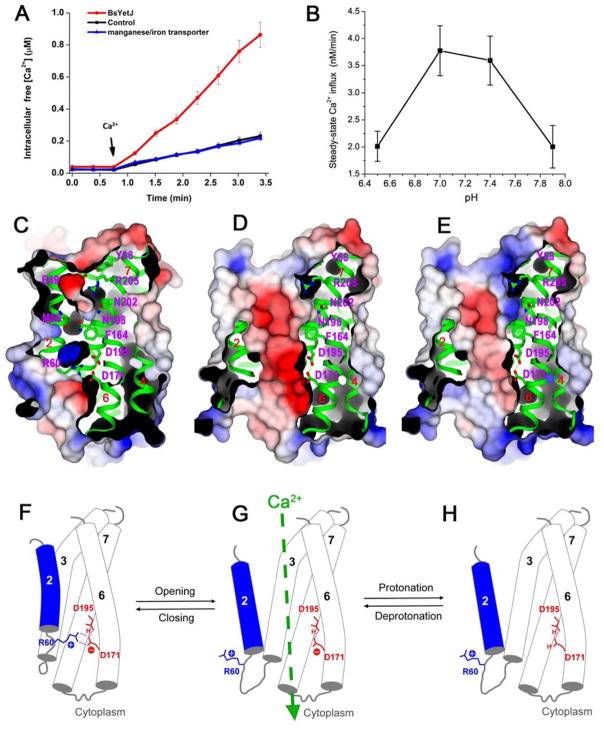
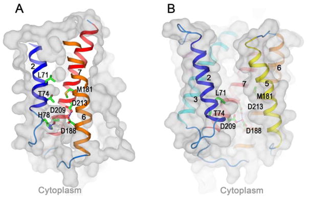
Calcium homeostasis balances passive calcium leak and active calcium uptake. Human Bax inhibitor-1 (hBI-1) is an antiapoptotic protein that mediates a calcium leak and is representative of a highly conserved and widely distributed family, the transmembrane Bax inhibitor motif (TMBIM) proteins. Here, we present crystal structures of a bacterial homolog and characterize its calcium leak activity. The structure has a seven-transmembrane-helix fold that features two triple-helix sandwiches wrapped around a central C-terminal helix. Structures obtained in closed and open conformations are reversibly interconvertible by change of pH. A hydrogen-bonded, pKa (where Ka is the acid dissociation constant)-perturbed pair of conserved aspartate residues explains the pH dependence of this transition, and biochemical studies show that pH regulates calcium influx in proteoliposomes. Homology models for hBI-1 provide insights into TMBIM-mediated calcium leak and cytoprotective activity.


Figures

Fig. 1
Fig. 1. Structures of BsYetJ
(A) Ribbon drawing of form-1 structure determined at pH 8. (B) Ribbon drawing of form-2 structure determined at pH 6. The views of (A) and (B) are from the membrane, and each structure has its seven TM helices color-coded. All connecting loops are colored in gray. (C) A cylinder diagram of the form-2 structure to show the structural features from a periplasmic view. The coloring is as for (B).
Fig. 2
Fig. 2. Structural features
(AB) Two triple-helix sandwich substructures consist of TM1-3 (A) and TM4-6 (B). The color scheme is as Fig. 1B. Insets in (A) and (B) are, respectively, the close α-helical contacts between TM1 and TM3 and between TM4 and TM6. Cα-H······O contacts between 2.3 Å - 3.5 Å were drawn as magenta dashes. (C) Overall pseudo-inverse symmetry with the triple-helix sandwiches in magenta for TM1-3 and green for TM4-6. The pseudo twofold axis is on the middle of the red TM7. (D) Superimposition of the two symmetric components.
Fig. 3
Fig. 3. Pore opening and closing regulated by pH
(A–B) Superimposition of the two conformationally different structures at pH 8 (cyan and blue) and at pH 6 (gray and magenta). (A) is cytoplasmic view and (B) is membrane view. (C) Electron density of the pH 7 structure showing alternative conformations of TM2. The 2Fo-Fc electron densities were drawn as gray isomeshes at 0.8s. The side chains of TM2 in closed and open conformations were drawn respectively as blue and magenta sticks. (D) Overall pH 7 structure with two alternative conformations. Deviated side chains are shown as sticks: cyan for closed conformation and gray for open conformation.
Fig. 4
Fig. 4. Di-aspartyl pH sensor
(A) H-bond interactions within the pore of the closed-conformation structure. (BD) Successive structures from intra-crystalline transitions with superimposed 2Fo-Fc electron densities contoured at two levels, 1.2s (gray) and 3.0s (magenta). (B) Starting form-1 structure at pH 8. (C) Structure after soaking a form-1 crystal into a medium at pH 6, disrupting the interactions between Arg60 and Asp171. (D) Structure after reversal, from first soaking at pH 6 and then back-soaking to pH 8, thereby reclosing the pore and restoring the interactions between Arg60 and Asp171.
Fig. 5
Fig. 5. Structural and functional characterization of calcium leak
(A) Calcium influx into bacteria over-expressing BsYetJ. An empty plasmid and a manganese transporter were used as negative controls. Error bars indicate the standard error of the mean (±SEM, n=9). (B) Calcium influx into proteoliposomes. Error bars indicate the standard error of the mean (±SEM, n=3). (C) Electrostatic surface of the closed-conformation structure showing charged surface concavities and internal cavities, but a blocked pore. (D) Electrostatic surface of the open-conformation structure at pH 7.4 where the cleft is electronegative. (E) Electrostatic surface for the open-conformation structure at pH 6 where the cleft is more neutral. The contour level of the electrostatics surface is at ±5 kT/e. Red, negative potential; blue, positive potential. (F–H) A proposed model for pH-sensitive calcium leak. (F) At higher pH (e.g. 8), Asp195 is protonated and Asp171 is deprotonated. Asp171 forms two H-bonds with positively charged Arg60 and the Arg60/Asp171 latch closes the pore. (G) When in the open conformation at a more neutral pH (e.g. 7.4), Asp171 may equilibrate between protonated and deprotonated states. Calcium passage occurs only when Asp171 is transiently deprotonated. (H) At lower pH (e.g. 6), the equilibration will favor more complete protonation of Asp171, disfavoring calcium passage due to pore neutralization. Cartoons (F–H) correspond to structures (C–E) directly above.
Fig. 6
Fig. 6. Homology models of human Bax inhibitor 1 (hBI-1)
(AB) Homology models of hBI-1 in its closed conformation (A) and open conformation (B). The conserved di-aspartyl Asp188/Asp213 unit is shown within the membrane in each case, and a third aspartate, Asp209, is shown interacting with His78 in (A) and with Asp188 in (B). In the closed-conformation (A), the indicated pore-sealing residues, Leu71, Thr74 and Met181, separate concave surfaces invaginating from opposite membrane face.

References

    1. Berridge MJ, Bootman MD, Roderick HL. Calcium signalling: dynamics, homeostasis and remodelling. Nat Rev Mol Cell Biol. 2003;4:517–529. - PubMed
    1. Smaili SS, Pereira GJ, Costa MM, Rocha KK, Rodrigues L, et al. The role of calcium stores in apoptosis and autophagy. Curr Mol Med. 2013;13:252–265. - PubMed
    1. Orrenius S, Zhivotovsky B, Nicotera P. Regulation of cell death: the calcium-apoptosis link. Nat Rev Mol Cell Biol. 2003;4:552–565. - PubMed
    1. Scorrano L, Oakes SA, Opferman JT, Cheng EH, Sorcinelli MD, et al. BAX and BAK regulation of endoplasmic reticulum Ca2+: a control point for apoptosis. Science. 2003;300:135–139. - PubMed
    1. Palmer AE, Jin C, Reed JC, Tsien RY. Bcl-2-mediated alterations in endoplasmic reticulum Ca2+ analyzed with an improved genetically encoded fluorescent sensor. Proc Natl Acad Sci USA. 2004;101:17404–17409. - PMC - PubMed

Publication types