. 2014 Jun 6;344(6188):1160-1164.
doi: 10.1126/science.1250778.

A mutually assured destruction mechanism attenuates light signaling in Arabidopsis

Affiliations

A mutually assured destruction mechanism attenuates light signaling in Arabidopsis

Weimin Ni et al. Science. .

Abstract

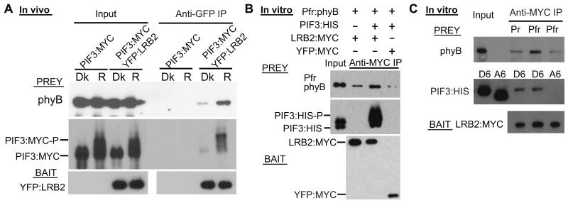
After light-induced nuclear translocation, phytochrome photoreceptors interact with and induce rapid phosphorylation and degradation of basic helix-loop-helix transcription factors, such as PHYTOCHROME-INTERACTING FACTOR 3 (PIF3), to regulate gene expression. Concomitantly, this interaction triggers feedback reduction of phytochrome B (phyB) levels. Light-induced phosphorylation of PIF3 is necessary for the degradation of both proteins. We report that this PIF3 phosphorylation induces, and is necessary for, recruitment of LRB [Light-Response Bric-a-Brack/Tramtrack/Broad (BTB)] E3 ubiquitin ligases to the PIF3-phyB complex. The recruited LRBs promote concurrent polyubiqutination and degradation of both PIF3 and phyB in vivo. These data reveal a linked signal-transmission and attenuation mechanism involving mutually assured destruction of the receptor and its immediate signaling partner.


Figures

Fig. 1
Fig. 1
Light-induced phosphorylation of PIF3 promotes interaction with Cullin E3-ligase substrate-recognition subunit LRB2. (A) Light induces LRB2 and PIF3 interaction in vivo. Dark-grown seedlings from either PIF3:MYC or PIF3:MYC/YFP:LRB2 double-transgenic lines were pretreated with MG132, then either kept in the dark (Dk) or irradiated with a pulse of red light (Rp) before protein extraction and co-immunoprecipitation (Co-IP) with an anti-GFP antibody. The proteins were analyzed by Western blot with either anti-MYC antibody (Prey, Top and middle panels), or anti-GFP antibody (Bait, Bottom panel). (B –D) Phosphorylation or phosphomimic mutations of the light-induced phosphosite residues in PIF3 promote its association with LRB2 in vitro. (B) and (C) In vitro-expressed recombinant LRB2 or YFP-control (Bait) proteins were precipitated with a MYC antibody, then incubated with normal (WT) PIF3 or the indicated light-induced-phosphosite mutant variants (A6, A20, D6, D19) of PIF3 (Prey). Bait and prey proteins were detected by Western blot using anti-MYC and anti-His antibody, respectively. All proteins were expressed in Hela cell lysate. A: Ser/Thr to Ala substitutions; D: Ser/Thr to Asp substitutions. A6/D6: PIF3 with substitutions in the six strongly light-induced phosphosite residues (8) (see Fig. S4A). A20/D19: PIF3 with substitutions in the majority of the light-induced phosphosite residues. Co-IP products were treated with alkaline phosphatase (CIAP) in panel (B). (D) PIF3-A6, PIF3-WT or PIF3-D6 expressed in Hela cell lysates were either treated (+) or not (−) with CIAP or with heat inactivated CIAP before Western blot analysis using anti-His antibody. (E) High affinity interaction of LRB2 and PIF3 in vitro requires multisite light-induced phosphorylation of PIF3. Immunoprecipitations and Western blots as in (C). PIF3 mutant-variant substitutions: D1: S88D, D3: S58/S88/S102D. See Fig. S4A for all S to A or D substitutions.
Fig. 2
Fig. 2
Light-induced phosphorylation of PIF3 promotes phyB-LRB2 association. (A) Light induces phyB-LRB2 association in vivo. Proteins were extracted and immunoprecipitated using the same lines and procedure as in Fig. 1A, and then subjected to Western blot analysis using anti-phyB antibody (Prey, top panel), anti-MYC antibody (Prey, middle panel), or anti-GFP antibody (Bait, bottom panel). (B) Phosphorylated PIF3 promotes phyB-LRB2 association in vitro. In vitro-expressed recombinant LRB2:MYC or YFP:MYC-control bait proteins were incubated with phyB-Pfr in the presense (+) or absence (−) of PIF3:His prey proteins, and immunoprecipitated with anti-MYC antibody. Western blots were probed with anti-phyB (Top), anti-His-epitope (middle) or anti-MYC (bottom) antibodies. (C) PIF3-D6 phosphomimic (8) (see Fig. S4A) promotes Pfr dependent phyB-LRB2 association in vitro. In vitro-expressed recombinant LRB2:MYC bait protein was incubated with phyB (as Pr or Pfr) and PIF3:His (D6- or A6-mutant (8) (see Fig. S4A)) prey proteins, and immunoprecipitated with anti-MYC antibody. Western blots as in (B).
Fig. 3
Fig. 3
LRBs function in both PIF3 and PIF3-mediated-phyB degradation in the light. (A) Endogenous PIF3 degradation is reduced in the lrb1lrb2lrb3 triple-mutant (lrb123) compared to Col wild-type (WT) seedlings in the light. Dark-grown seedlings of Col and lrb123 were irradiated with red light (R) for the period indicated before protein extraction and Western blot analysis using anti-PIF3 antibody (Left panel). PIF3: unphosphorylated PIF3; PIF3-P: phosphorylated PIF3; NS: nonspecific bands. Right panel shows quantification of the Western blot results from both the Left panel (blue and red curves) and Fig. S6B (green and purple curves) after normalization to the Tubulin loading control. The dark PIF3 level was set as 100%. (B) PIF3:GFP accumulates in the phosphorylated form in the lrb123 triple mutant in the light. Seedling growth was as in panel (A). Extracted proteins were either analyzed directly (Top two panels), or after phosphatase (CIAP) treatment (Third panel) by Western blot using anti-GFP antibody. (C) LRBs are essential for light-induced, PIF3-promoted feedback degradation of phyB. Light-induced degradation of phyB (accelerated by PIF3:GFP overexpression) is absent in the lrb123 mutant background. Dark-grown seedlings of the indicated genotypes were irradiated with 3 (R3h), 24 (R24h) or 96 (Rc96h) hours of continuous red light before protein extraction and Western blot analysis using an anti-phyB antibody (Left and middle panels). Tubulin was used as a loading control. Right panel shows quantification of the relative phyB/Tubulin protein levels from left and middle panels. Dark levels were set as 100%. (D) PIF3:GFP over-expression does not complement the short-hypocotyl phenotype of the lrb123 mutant in the light. Seedlings of the indicated genotypes were grown for 4 days in the dark (Dk) or continuous red light (Rc). Visible phenotypes (left); hypocotyl lengths (right). Error bars represent standard errors. (E) Endogenous PIF3 levels are strongly reduced in dark-grown seedlings of the cop1 and spa123 mutants.
Fig. 4
Fig. 4
LRB E3 ligases function in polyubiquitination of both PIF3 and phyB. (A) Light-induced poly-ubiquitination of PIF3 is reduced in the lrb123 mutant. Dark-grown wild-type (col) or lrb123-mutant seedlings, transgenically, or not, expressing PIF3:GFP (PIF3:GFP/col and PIF3:GFP/lrb123, respectively), were irradiated with 10 min of red light (R), or not (Dk), before protein extraction and immunoprecipitation (IP) using anti-GFP antibody. The IP products were then analyzed by Western blot with anti-GFP antibody (left panel) or anti-ubiquitin antibody (right panel). (B) Light-induced mobility shift of PIF3-bound phyB (phyB-ubi) is absent in the lrb123 mutant. Co-IP products prepared as in panel A were analyzed with anti-phyB antibody. (C) Light-induced poly-ubiquitination of both phyB and PIF3 is strongly reduced in the lrb123 mutant. Seedling growth and extraction was as in panel A. Total ubiquitinated proteins were immunoprecipitated with TUBEs (Tandem Ubiquitin Binding Entities), then analyzed by Western blot using antibody against ubiquitin (left panel), phyB (middle panel), or GFP (right panel). (D) A Cullin3-LRB2 E3 ubiquitin-ligase complex ubiquitinates phosphomimic PIF3 (D6) in vitro. A Cullin3 E3 ubiquitin ligase complex was assembled with recombinant LRB2 protein and incubated with D6 or A20 PIF3:MYC (8) (see Fig. S4A for D6) variants in vitro (see Methods) before western blot analysis with anti-MYC antibody.

References

    1. Avraham R, Yarden Y. Feedback regulation of EGFR signalling: decision making by early and delayed loops. Nat Rev Mol Cell Biol. 2011;12:104. - PubMed
    1. Yarden Y. The biological framework: translational research from bench to clinic. Oncologist. 2010;15:1. - PubMed
    1. Amit I, et al. A module of negative feedback regulators defines growth factor signaling. Nat Genet. 2007;39:503. - PubMed
    1. Leivar P, Quail PH. PIFs: pivotal components in a cellular signaling hub. Trends Plant Sci. 2011;16:19. - PMC - PubMed
    1. Jiao Y, Lau OS, Deng XW. Light-regulated transcriptional networks in higher plants. Nat Rev Genet. 2007;8:217. - PubMed

Publication types

MeSH terms