

Stress-induced mast cell activation in glabrous and hairy skin
- PMID: 24904196
- PMCID: PMC4034722
- DOI: 10.1155/2014/105950
Stress-induced mast cell activation in glabrous and hairy skin
Abstract
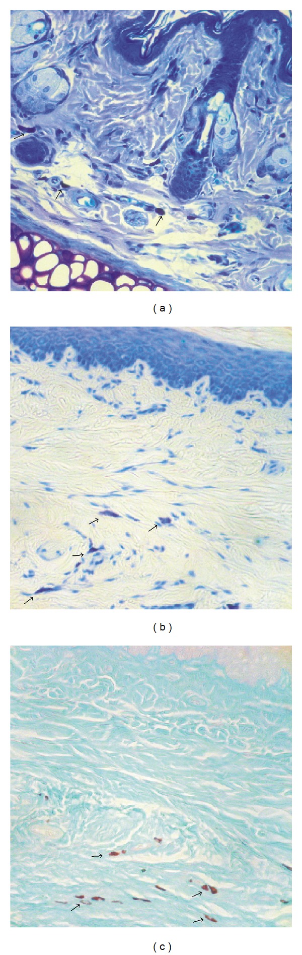
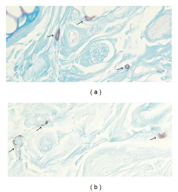
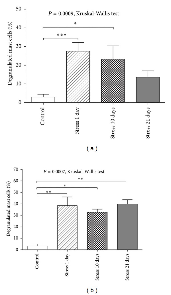
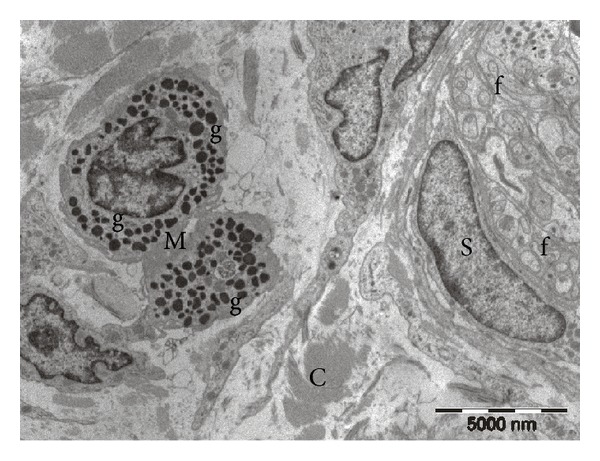
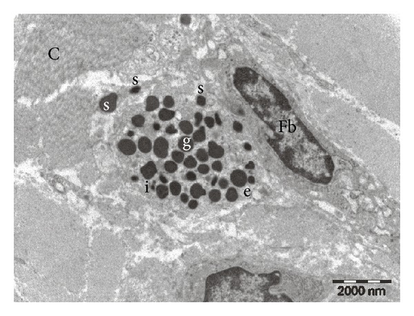
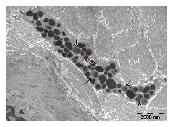
Mast cells play a key role in modulation of stress-induced cutaneous inflammation. In this study we investigate the impact of repeated exposure to stress on mast cell degranulation, in both hairy and glabrous skin. Adult male Wistar rats were randomly divided into four groups: Stress 1 day (n = 8), Stress 10 days (n = 7), Stress 21 days (n = 6), and Control (n = 8). Rats in the stress groups were subjected to 2 h/day restraint stress. Subsequently, glabrous and hairy skin samples from animals of all groups were collected to assess mast cell degranulation by histochemistry and transmission electron microscopy. The impact of stress on mast cell degranulation was different depending on the type of skin and duration of stress exposure. Short-term stress exposure induced an amplification of mast cell degranulation in hairy skin that was maintained after prolonged exposure to stress. In glabrous skin, even though acute stress exposure had a profound stimulating effect on mast cell degranulation, it diminished progressively with long-term exposure to stress. The results of our study reinforce the view that mast cells are active players in modulating skin responses to stress and contribute to further understanding of pathophysiological mechanisms involved in stress-induced initiation or exacerbation of cutaneous inflammatory processes.
Figures
References
-
- Neagu M, Constantin C, Tanase C. Immune-related biomarkers for diagnosis/prognosis and therapy monitoring of cutaneous melanoma. Expert Review of Molecular Diagnostics. 2010;10(7):897–919. - PubMed
-
- Ardigò M, Longo C, Cristaudo A, Berardesca E, Pellacani G. Evaluation of allergic vesicular reaction to patch test using in vivo confocal microscopy. Skin Research and Technology. 2012;18(1):61–63. - PubMed
-
- Koo J, Lebwohl A. Psychodermatology: the mind and skin connection. The American Family Physician. 2001;64(11):1873–1878. - PubMed
-
- Arck P, Paus R. From the brain-skin connection: the neuroendocrine-immune misalliance of stress and itch. NeuroImmunoModulation. 2007;13(5-6):347–356. - PubMed
-
- Tausk F, Elenkov I, Moynihan J. Psychoneuroimmunology. Dermatologic Therapy. 2008;21(1):22–31. - PubMed
Publication types
MeSH terms
LinkOut - more resources
Full Text Sources
Other Literature Sources
