

One-pot synthesis of magnetic nanoclusters enabling atherosclerosis-targeted magnetic resonance imaging
- PMID: 24904209
- PMCID: PMC4039422
- DOI: 10.2147/IJN.S55525
One-pot synthesis of magnetic nanoclusters enabling atherosclerosis-targeted magnetic resonance imaging
Abstract
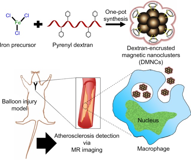
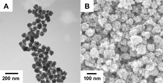
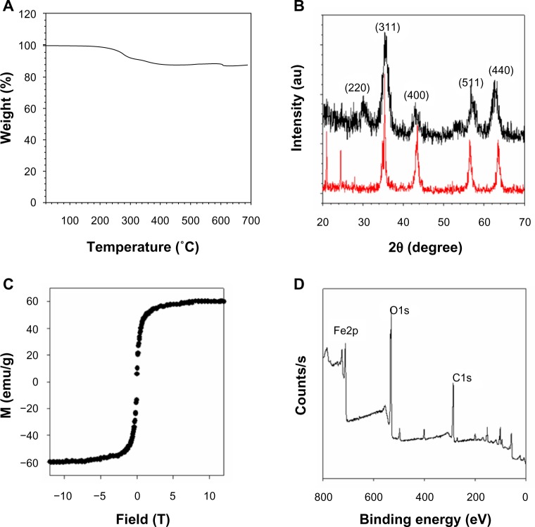
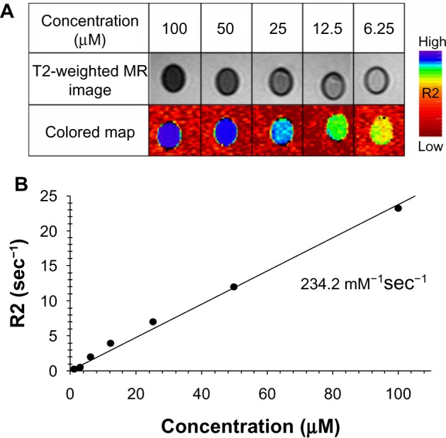
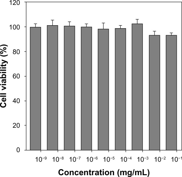
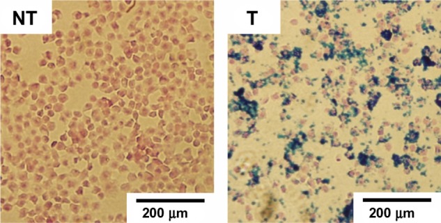
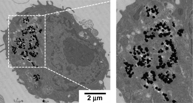
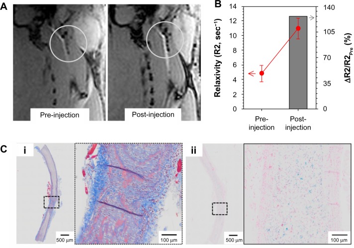
In this study, dextran-encrusted magnetic nanoclusters (DMNCs) were synthesized using a one-pot solution phase method for detection of atherosclerosis by magnetic resonance imaging. Pyrenyl dextran was used as a surfactant because of its electron-stabilizing effect and its amphiphilic nature, rendering the DMNCs stable and water-dispersible. The DMNCs were 65.6±4.3 nm, had a narrow size distribution, and were superparamagnetic with a high magnetization value of 60.1 emu/g. Further, they showed biocompatibility and high cellular uptake efficiency, as indicated by a strong interaction between dextran and macrophages. In vivo magnetic resonance imaging demonstrated the ability of DMNCs to act as an efficient magnetic resonance imaging contrast agent capable of targeted detection of atherosclerosis. In view of these findings, it is concluded that DMNCs can be used as magnetic resonance imaging contrast agents to detect inflammatory disease.
Keywords: atherosclerosis; dextran; macrophages; magnetic nanocrystal; magnetic resonance imaging.
Figures
References
-
- Jun YW, Huh YM, Choi JS, et al. Nanoscale size effect of magnetic nanocrystals and their utilisation for cancer diagnosis via magnetic resonance imaging. J Am Chem Soc. 2005;127(16):5732–5733. - PubMed
-
- Huh YM, Jun YW, Song HT, et al. In vivo magnetic resonance detection of cancer by using multifunctional magnetic nanocrystals. J Am Chem Soc. 2005;127(35):12387–12391. - PubMed
-
- Jun YW, Seo JW, Cheon J. Nanoscaling laws of magnetic nanoparticles and their applicabilities in biomedical sciences. Acc Chem Res. 2008;41(2):179–189. - PubMed
-
- Lee JH, Huh YM, Jun YW, et al. Artificially engineered magnetic nanoparticles for ultra-sensitive molecular imaging. Nat Med. 2007;13(1):95–99. - PubMed
-
- Lu J, Ma S, Sun J, et al. Manganese ferrite nanoparticle micellar nanocomposites as MRI contrast agent for liver imaging. Biomaterials. 2009;30(15):2919–2928. - PubMed
Publication types
MeSH terms
Substances
LinkOut - more resources
Full Text Sources
Other Literature Sources
Medical
