. 2014 May 19:8:87.
doi: 10.3389/fnins.2014.00087. eCollection 2014.

Closed-loop control of spinal cord stimulation to restore hand function after paralysis

Affiliations

Closed-loop control of spinal cord stimulation to restore hand function after paralysis

Jonas B Zimmermann et al. Front Neurosci. .

Abstract

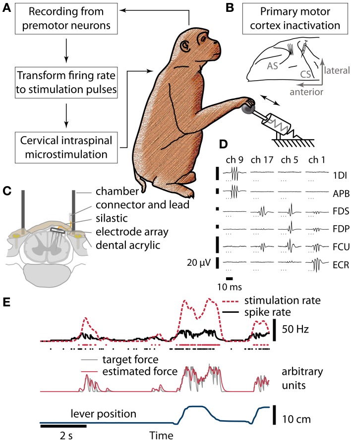
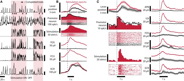
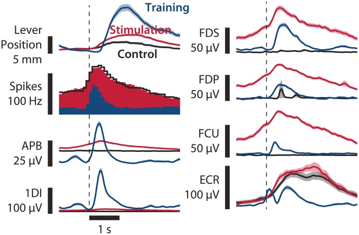
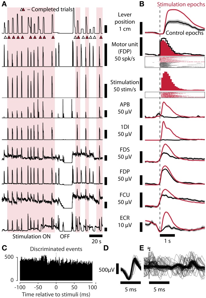
As yet, no cure exists for upper-limb paralysis resulting from the damage to motor pathways after spinal cord injury or stroke. Recently, neural activity from the motor cortex of paralyzed individuals has been used to control the movements of a robot arm but restoring function to patients' actual limbs remains a considerable challenge. Previously we have shown that electrical stimulation of the cervical spinal cord in anesthetized monkeys can elicit functional upper-limb movements like reaching and grasping. Here we show that stimulation can be controlled using cortical activity in awake animals to bypass disruption of the corticospinal system, restoring their ability to perform a simple upper-limb task. Monkeys were trained to grasp and pull a spring-loaded handle. After temporary paralysis of the hand was induced by reversible inactivation of primary motor cortex using muscimol, grasp-related single-unit activity from the ventral premotor cortex was converted into stimulation patterns delivered in real-time to the cervical spinal gray matter. During periods of closed-loop stimulation, task-modulated electromyogram, movement amplitude, and task success rate were improved relative to interleaved control periods without stimulation. In some sessions, single motor unit activity from weakly active muscles was also used successfully to control stimulation. These results are the first use of a neural prosthesis to improve the hand function of primates after motor cortex disruption, and demonstrate the potential for closed-loop cortical control of spinal cord stimulation to reanimate paralyzed limbs.

Keywords: brain-computer interface; closed-loop neuro-prosthetics devices; electrical stimulation; grasp; hand movements; intraspinal microstimulation; non-human primate model; spinal cord injury.


Figures

Figure 1
Figure 1
Brain control of spinal cord stimulation after reversible paralysis. (A) Schematic of the closed-loop stimulation protocol. Spiking activity recorded from neurons in PMv was converted in real-time to stimulation delivered to the spinal cord while monkeys performed a grasp-and-pull task. (B) Hand and forearm muscles were temporarily paralyzed by micro-injections of muscimol into hand area of M1, anterior to the central sulcus (CS). Neural activity was recorded from PMv, in the posterior bank of the arcuate sulcus (AS). (C) Cross-section of the spinal stimulation implant used in monkey R. (D) Sample EMG responses elicited by stimulation of different channels of the FMA (arranged in columns, channels 1, 5, 9, and 17 of the array chosen to illustrate different response properties; 3 biphasic pulses at 300 Hz, stimulation currents between 12 and 81 μA. 1DI, first dorsal interosseus; APB, abductor pollicis brevis; FDS, flexor digitorum superficialis; FDP, flexor digitorum profundus; FCU, flexor carpi ulnaris; ECR, extensor carpi radialis). (E) From neural spikes to stimulation pulses (simulated data). Spikes (black dots) were used to estimate instantaneous firing rate (black line) in real time. The firing rate was transformed to yield a target force function (gray line, arbitrary units). Whenever the estimated stimulation-induced force (red line, a. u.) was below the target, a stimulus pulse (red dots) was delivered. Smoothed stimulation rate (red broken line) and lever position (blue line) are shown for comparison.
Figure 2
Figure 2
Brain-controlled spinal stimulation improves task performance and restores muscle activity. (A) Lever position, neural firing rate, stimulation rate, and EMG recorded from FDS, FCU, and ECR recorded during a brain-controlled stimulation session (monkey B). Consecutive stimulation epochs (shaded) and control epochs with no stimulation (no shading) are shown, incorporating several successful trials (indicated by triangles, filled: stimulation, open: control). (B) Average data from stimulation (124 trial attempts) and control (73 trial attempts) epochs aligned to attempt onset (inferred from neural firing rate exceeding 90 spikes/s). Raster plots show 20 stimulation and 20 control trial attempts. Shaded areas indicate standard error of the mean (SEM). Monkey B, session B100711000. (C) Similar to (B), monkey R. Trials aligned to PMv neuron firing rate exceeding 11 Hz. One hundred twenty-nine stimulation trials and 35 control trials over a period of 29 min are shown. Shaded areas: SEM. Session Rv110719002.
Figure 3
Figure 3
Comparison of paralysis+stimulation, paralysis, and training trials. Averages of lever position, neural firing, and EMG of various hand and forearm muscles are shown. Paralysis+stimulation (red, n = 124 trials) and paralysis (black, n = 73 trials) epochs are the same as in Figure 2. Training epochs (blue, n = 163 trials) are from a session 3 days before in which no muscimol was injected. Epochs are aligned to threshold crossing of neural firing rate (90 Hz for paralysis session, 40 Hz for training session, broken vertical line). Muscle activation due to spinal stimulation in finger flexors FDS and FDP and wrist flexor FCU resembles normal activation in shape and amplitude. Monkey B, sessions B100708001 and B100711000. Shaded areas: SEM.
Figure 4
Figure 4
Task performance is restored by spinal stimulation controlled by residual muscle activity. (A) Lever position, motor unit firing rate, stimulation rate, and EMG recorded from APB, 1DI, FDS, FDP, FCU, and ECR, during a session in which residual motor unit activity from FDP was used to control stimulation (monkey R). (B) Average data from stimulation (178 trial attempts) and control (43 trial attempts) epochs aligned to attempt onset (inferred from motor unit firing rate exceeding 20 Hz). Shaded areas: SEM. Session Rv110714003. (C) Peri-Stimulus Time Histogram showing discriminated motor unit action potentials used to control stimulation relative to occurrence of stimulus pulses (0 ms). The absence of a peak following the stimulus indicates that the discriminator was neither triggered by a stimulus artifact nor by a stimulus-evoked motor response (which could lead to a positive feed-back loop). (D) Sample raw discriminated FDP motor unit potentials (n = 100). (E) Raw FDP EMG traces aligned to stimulation events at vertical bar (n = 100).
Figure 5
Figure 5
Development of motor thresholds over time. Dots show measured stimulation thresholds for individual electrodes of monkey R's array implant over the course of the experiment. Dots are color-coded for length of the electrode. The solid red line shows the average thresholds for each session (shaded area, SEM). Dashed lines represent linear fits for the three lengths of electrodes (gray), and all electrodes (red). The cartoon (right) shows the distribution of electrode lengths over the array and its position within the cord.
Figure 6
Figure 6
Development of mean firing rate of the neuron used to control spinal stimulation over the course of the experiment in monkey B. We used the same neuron to control spinal stimulation over the course of 18 days. Mean firing rate was higher during stimulation sessions (solid symbols) than in training sessions (open symbols), and increased over time (see Table 1 for linear regression statistics).

References

    1. Bamford J. A., Putman C. T., Mushahwar V. K. (2005). Intraspinal microstimulation preferentially recruits fatigue-resistant muscle fibers and generates gradual force in rat. J. Physiol. 569, 873–884 10.1113/jphysiol.2005.094516 - DOI - PMC - PubMed
    1. Bansal A. K., Truccolo W., Vargas-Irwin C. E., Donoghue J. P. (2012). Decoding 3D reach and grasp from hybrid signals in motor and premotor cortices: spikes, multiunit activity, and local field potentials. J. Neurophysiol. 107, 1337–1355 10.1152/jn.00781.2011 - DOI - PMC - PubMed
    1. Collinger J. L., Wodlinger B., Downey J. E., Wang W., Tyler-Kabara E. C., Weber D. J., et al. (2013). High-performance neuroprosthetic control by an individual with tetraplegia. Lancet 381, 557–564 10.1016/S0140-6736(12)61816-9 - DOI - PMC - PubMed
    1. Craggs M. D. (1975). Cortical control of motor prostheses: using the cord-transected baboon as the primate model for human paraplegia. Adv. Neurol. 10, 91–101 - PubMed
    1. DeVivo M. J. (2012). Epidemiology of traumatic spinal cord injury: trends and future implications. Spinal Cord 50, 365–372 10.1038/sc.2011.178 - DOI - PubMed