Review
. 2014 May 27:7:48.
doi: 10.3389/fnmol.2014.00048. eCollection 2014.

Cellular function and pathological role of ATP13A2 and related P-type transport ATPases in Parkinson's disease and other neurological disorders

Affiliations
Review

Cellular function and pathological role of ATP13A2 and related P-type transport ATPases in Parkinson's disease and other neurological disorders

Sarah van Veen et al. Front Mol Neurosci. .

Abstract

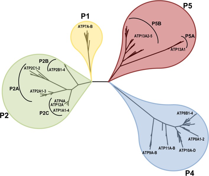
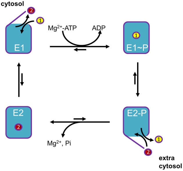
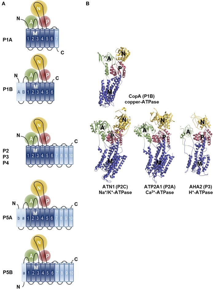
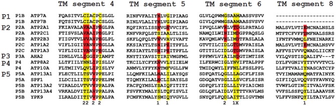
Mutations in ATP13A2 lead to Kufor-Rakeb syndrome, a parkinsonism with dementia. ATP13A2 belongs to the P-type transport ATPases, a large family of primary active transporters that exert vital cellular functions. However, the cellular function and transported substrate of ATP13A2 remain unknown. To discuss the role of ATP13A2 in neurodegeneration, we first provide a short description of the architecture and transport mechanism of P-type transport ATPases. Then, we briefly highlight key P-type ATPases involved in neuronal disorders such as the copper transporters ATP7A (Menkes disease), ATP7B (Wilson disease), the Na(+)/K(+)-ATPases ATP1A2 (familial hemiplegic migraine) and ATP1A3 (rapid-onset dystonia parkinsonism). Finally, we review the recent literature of ATP13A2 and discuss ATP13A2's putative cellular function in the light of what is known concerning the functions of other, better-studied P-type ATPases. We critically review the available data concerning the role of ATP13A2 in heavy metal transport and propose a possible alternative hypothesis that ATP13A2 might be a flippase. As a flippase, ATP13A2 may transport an organic molecule, such as a lipid or a peptide, from one membrane leaflet to the other. A flippase might control local lipid dynamics during vesicle formation and membrane fusion events.

Keywords: alpha-synuclein; dystonia; flippase; heavy metal toxicity; lysosome; mitochondria; mitophagy; parkinsonism.


Figures

Figure 1
Figure 1
Phyologenetic tree of the human P-type ATPases. Phylogenetic tree based on the core protein sequences of 137 animal homologues of the 36 human P-type ATPase isoforms. ATP13A2 homologues were obtained from the database Homologene http://www.ncbi.nlm.nih.gov/homologene. Core protein sequences were generated according to the methodology described in Axelsen and Palmgren (1998). The 36 human P-type ATPases are indicated. Of note, only animal isoforms are depicted, so the P3A-type ATPases, which are uniquely found in fungi and plants and the small class of bacterial Mg+-ATPases of the P3B group and bacterial pumps belonging to P1A are not represented. The phylogenetic tree was rendered using www.phylogeny.fr (Dereeper et al., 2008, 2010).
Figure 2
Figure 2
General Post-Albers reaction scheme for P-type ATPases. A cytosolic ligand (yellow, transported ligand 1) is transported to the extracytosolic space, whereas an extracytosolic ligand (red, counter-transported ligand 2) is imported into the cytosol. Note that the number of ligands in each direction may vary between different P-type ATPase isoforms. In short, P-type ATPases switch between two major conformations E1, with ligand binding sites facing the cytosol, and E2, with ligand binding sites facing the extracytosolic side of the membrane. The induced fit of ligand 1 binding in E1 promotes phosphorylation by Mg+-ATP. In this E1~P state the ligand 1 becomes occluded. The rate-limiting E1~P to E2-P transition is accompanied by major conformational changes, reorienting the ligand-binding sites toward the extracytosolic space. This decreases the affinity of the binding site for ligand 1, whereas the affinity for ligand 2 is increased. As a result, ligand 1 is released into the extracytosolic space via an open exit pathway for ligand 1 and the counter-transported ligand 2 can enter the binding cavity. The resulting conformational changes lead to dephosphorylation of E2P and the released inorganic phosphate is expelled. The ligand 2 becomes occluded, whereupon the pump is reset to the E1 state, reducing the affinity for ligand 2. The pump can now start a new cycle.
Figure 3
Figure 3
Topology and architecture of the catalytic subunits of P-type ATPases. (A) Planar topology models of the five classes of P-type ATPases (P1-P5). Nucleotide-binding domains (N, yellow), actuator domains (A, green) and phosphorylation domains (P, red) are indicated. The 6 TM helices (Polymeropoulos et al., ; Spillantini et al., ; Jankovic, ; Lees et al., ; Auluck et al., ; Tolleson and Fang, 2013) form the core segment of the membrane (M) domain of all P-type ATPases, which is depicted in dark blue, whereas additional N- and C-terminal helices are shown in light blue. Of note, there is one exception for the P2 ATPases, a splice variant of ATP2A2, SERCA2b, harbors an 11th TM helix at the C-terminus (not shown, Vandecaetsbeek et al., 2009). (B) Resolved P-type ATPase crystal structures, known up until now. The Legionella pneumophila CopA copper-ATPase (PDB 3RFU), a P1B-type ATPase; the rabbit P2A-type ATPase ATP2A1 (SERCA1a, PDB 2AGV), the Squalus acanthias Na+/K+-ATPase α-subunit ATN1 (PDB 3A3Y), a member of the P2C group and the Arabidopsis thaliana proton pump AHA2 (PBD 3B8C) of the P3-type ATPases. N-, A-, P- and M-domains are indicated with similar colors as in the planar models. Note that the obligatory subunits of the P1A, P2C and P4 are not shown.
Figure 4
Figure 4
Sequence comparison of the TM helices in P-type ATPases of various subfamilies. The residues involved in Ca2+ binding in the two Ca2+ binding sites (site 1 and 2) in the SERCA1a Ca2+ pump (ATP2A1) are distributed over four TM helices: M4, 5, 6 and 8. The colored residues are part of the Ca2+ binding sites in ATP2A1 and numbers 1 and 2 refer to the number of the Ca2+-binding site to which the residue contributes (x is contributing to both site 1 and site 2). The sequence of the M4, M5, M6 and M8 helices is compared with those of the P-type ATPases that are involved in neurological disorders. Also the yeast P5 ATPases Spf1p and Ypk9p, the Ca2+/Mn2+-ATPase SPCA as well as the proton pump AHA2 are included for comparison. M4 shows the highest degree of conservation. Highlighted in red are conserved residues as compared to the ATP2A1 Ca2+ binding site sequence, whereas in yellow the non-conserved residues are indicated. For each subfamily, a signature motif can be recognized in M4, which corresponds well with the substrate specificity. The PPELP and PPALP sequences of P5A- and P5B-type ATPases have little in common with other P-type ATPase signature motifs, which might indicate that the transported ligand is significantly different.
Figure 5
Figure 5
Genetic interactions of YPK9 in S. cerevisiae grouped according to cellular localization and function. Summary of the known genetic interactions of the yeast ATP13A2 ortholog YPK9. The data were collected from the yeast genome database (http://yeastgenome.org, see Supplemental Table 1) and were classified using BioGrid (http://thebiogrid.org) according to organellar distribution and function.

References

    1. Albers R. W. (1967). Biochemical aspects of active transport. Annu. Rev. Biochem. 36, 727–756 10.1146/annurev.bi.36.070167.003455 - DOI - PubMed
    1. Ambrosini A., D'Onofrio M., Grieco G. S., Di Mambro A., Montagna G., Fortini D., et al. (2005). Familial basilar migraine associated with a new mutation in the ATP1A2 gene. Neurology 65, 1826–1828 10.1212/01.wnl.0000187072.71931.c0 - DOI - PubMed
    1. Auluck P. K., Caraveo G., Lindquist S. (2010). alpha-Synuclein: membrane interactions and toxicity in Parkinson's disease. Annu. Rev. Cell Dev. Biol. 26, 211–233 10.1146/annurev.cellbio.042308.113313 - DOI - PubMed
    1. Auranen M., Vanhala R., Varilo T., Ayers K., Kempas E., Ylisaukko-Oja T., et al. (2002). A genomewide screen for autism-spectrum disorders: evidence for a major susceptibility locus on chromosome 3q25-27. Am. J. Hum. Genet. 71, 777–790 10.1086/342720 - DOI - PMC - PubMed
    1. Axelsen K. B., Palmgren M. G. (1998). Evolution of substrate specificities in the P-type ATPase superfamily. J. Mol. Evol. 46, 84–101 10.1007/PL00006286 - DOI - PubMed