. 2014 May 20:8:277.
doi: 10.3389/fnhum.2014.00277. eCollection 2014.

Evidence for evolutionary specialization in human limbic structures

Affiliations

Evidence for evolutionary specialization in human limbic structures

Nicole Barger et al. Front Hum Neurosci. .

Abstract

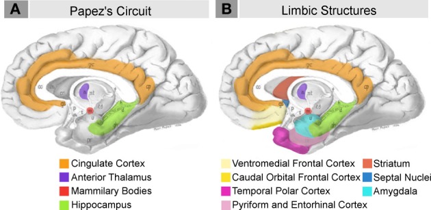
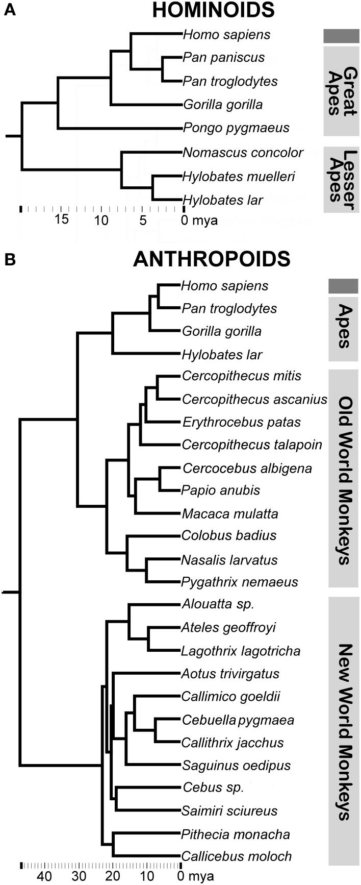
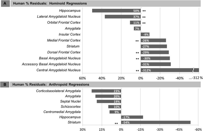
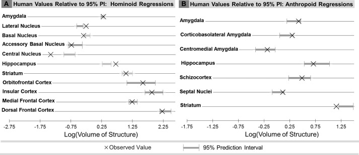
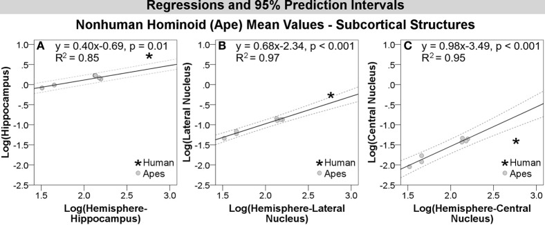
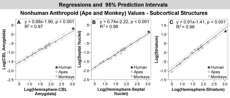
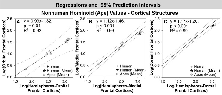
Increasingly, functional and evolutionary research has highlighted the important contribution emotion processing makes to complex human social cognition. As such, it may be asked whether neural structures involved in emotion processing, commonly referred to as limbic structures, have been impacted in human brain evolution. To address this question, we performed an extensive evolutionary analysis of multiple limbic structures using modern phylogenetic tools. For this analysis, we combined new volumetric data for the hominoid (human and ape) amygdala and 4 amygdaloid nuclei, hippocampus, and striatum, collected using stereological methods in complete histological series, with previously published datasets on the amygdala, orbital and medial frontal cortex, and insula, as well as a non-limbic structure, the dorsal frontal cortex, for contrast. We performed a parallel analysis using large published datasets including many anthropoid species (human, ape, and monkey), but fewer hominoids, for the amygdala and 2 amygdaloid subdivisions, hippocampus, schizocortex, striatum, and septal nuclei. To address evolutionary change, we compared observed human values to values predicted from regressions run through (a) non-human hominoids and (b) non-human anthropoids, assessing phylogenetic influence using phylogenetic generalized least squares regression. Compared with other hominoids, the volumes of the hippocampus, the lateral nucleus of the amygdala, and the orbital frontal cortex were, respectively, 50, 37, and 11% greater in humans than predicted for an ape of human hemisphere volume, while the medial and dorsal frontal cortex were, respectively, 26 and 29% significantly smaller. Compared with other anthropoids, only human values for the striatum fell significantly below predicted values. Overall, the data present support for the idea that regions involved in emotion processing are not necessarily conserved or regressive, but may even be enhanced in recent human evolution.

Keywords: amygdala; ape; comparative neuroanatomy; emotion; frontal cortex; hippocampus; hominoid; human brain evolution.


Figures

Figure 1
Figure 1
Schematic briefly summarizing neural systems proposed to process emotion, highlighting structures that are visible on the medial surface of the brain. Papez's (1937) original circuit (A) was expanded upon in the concept of the limbic system (B) to include a variety of subcortical and cortical territories (MacLean, ; Heimer and Van Hoesen, 2006). (Structures like the anterior insula and nucleus basalis of Meynert, which are not visible on the medial surface of the brain, are not represented here). Images modified from Papez's (1937) original drawing.
Figure 2
Figure 2
Consensus phylogeny for (A) hominoids and (B) anthropoids in the analysis with common classifications of major non-human primate clades indicated. Branch lengths represent evolutionary distance on the scale of millions of years (mya). Consensus phylogenies drawn from the 10 kTrees Website.
Figure 3
Figure 3
Human percent residual deviations from non-human regression lines for: (A) Human comparisons with other hominoids and (B) human comparisons with other anthropoids. Asterisks (**) indicate residuals that were significant, determined either by 95% prediction intervals (subcortical structures) or comparison of means at p < 0.05 (cortical structures).
Figure 4
Figure 4
Comparison of observed human brain structure volumes with the range of values predicted for human hemisphere volume from the 95% prediction intervals (PI) for: (A) Human comparisons with other hominoids and (B) human comparisons with other anthropoids.
Figure 5
Figure 5
Log-log regressions and 95% prediction intervals (dashed lines) of species average volumes (cc) for select subcortical structures in the hominoid dataset, which reached significance in statistical analyses, including: (A) hippocampus, (B) lateral amygdaloid nucleus, and (C) central amygdaloid nucleus.
Figure 6
Figure 6
Log-log regressions and 95% prediction intervals (dashed lines) of species average volumes (cc) for select structures in the anthropoid dataset, which reached significance or fell in the upper bounds of the prediction interval, including: (A) corticobasolateral amygdala, (B) septal nuclei, and (C) striatum. Abbr: CBL, corticobasolateral amygdala.
Figure 7
Figure 7
Log-log regressions and 95% prediction intervals (dashed lines) of species average volumes (cc) for select cortical structures in the hominoid dataset, which reached significance in statistical analyses, including: (A) orbital frontal cortex, (B) medial frontal cortex, and (C) dorsal frontal cortex.

References

    1. Adolphs R. (2009). The social brain: neural basis of social knowledge. Annu. Rev. Psychol. 60, 693–716 10.1146/annurev.psych.60.110707.163514 - DOI - PMC - PubMed
    1. Aldridge K. (2011). Patterns of differences in brain morphology in humans as compared to extant apes. J. Hum. Evol. 60, 94–105 10.1016/j.jhevol.2010.09.007 - DOI - PMC - PubMed
    1. Allman J. M., Tetreault N. A., Hakeem A. Y., Manaye K. F., Semendeferi K., Erwin J. M., et al. (2010). The von Economo neurons in frontoinsular and anterior cingulate cortex in great apes and humans. Brain Struct. Funct. 214, 495–517 10.1007/s00429-010-0254-0 - DOI - PubMed
    1. Amici F., Aureli F., Call J. (2008). Fission-fusion dynamics, behavioral flexibility, and inhibitory control in primates. Curr. Biol. 18, 1415–1419 10.1016/j.cub.2008.08.020 - DOI - PubMed
    1. Amunts K., Kedo O., Kindler M., Pieperhoff P., Mohlberg H., Shah N. J., et al. (2005). Cytoarchitectonic mapping of the human amygdala, hippocampal region and entorhinal cortex: intersubject variability and probability maps. Anat. Embryol. 210, 343–352 10.1007/s00429-005-0025-5 - DOI - PubMed