. 2014 May 26:5:79.
doi: 10.3389/fneur.2014.00079. eCollection 2014.

Insulin-Like Growth Factor Receptor Signaling is Necessary for Epidermal Growth Factor Mediated Proliferation of SVZ Neural Precursors in vitro Following Neonatal Hypoxia-Ischemia

Affiliations

Insulin-Like Growth Factor Receptor Signaling is Necessary for Epidermal Growth Factor Mediated Proliferation of SVZ Neural Precursors in vitro Following Neonatal Hypoxia-Ischemia

Dhivyaa Alagappan et al. Front Neurol. .

Abstract

In this study, we assessed the importance of insulin-like growth factor (IGF) and epidermal growth factor (EGF) receptor co-signaling for rat neural precursor (NP) cell proliferation and self-renewal in the context of a developmental brain injury that is associated with cerebral palsy. Consistent with previous studies, we found that there is an increase in the in vitro growth of subventricular zone NPs isolated acutely after cerebral hypoxia-ischemia; however, when cultured in medium that is insufficient to stimulate the IGF type 1 receptor, neurosphere formation and the proliferative capacity of those NPs was severely curtailed. This reduced growth capacity could not be attributed simply to failure to survive. The growth and self-renewal of the NPs could be restored by addition of both IGF-I and IGF-II. Since the size of the neurosphere is predominantly due to cell proliferation we hypothesized that the IGFs were regulating progression through the cell cycle. Analyses of cell cycle progression revealed that IGF-1R activation together with EGFR co-signaling decreased the percentage of cells in G1 and enhanced cell progression into S and G2. This was accompanied by increases in expression of cyclin D1, phosphorylated histone 3, and phosphorylated Rb. Based on these data, we conclude that coordinate signaling between the EGF receptor and the IGF type 1 receptor is necessary for the normal proliferation of NPs as well as for their reactive expansion after injury. These data indicate that manipulations that maintain or amplify IGF signaling in the brain during recovery from developmental brain injuries will enhance the production of new brain cells to improve neurological function in children who are at risk for developing cerebral palsy.

Keywords: cell proliferation; central nervous system; growth factors; regeneration; stem cell niche.


Figures
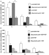
Figure 1
Figure 1
The increase in neurosphere number after neonatal H–I requires EGFR activation together with IGF-R co-signaling. SVZ cells were isolated from ipsilateral (IL), contralateral (CL), and sham-operated control (CTL) at 3-day recovery from H–I. The dissociated cells were cultured in media containing 25 μg/ml “high” insulin or 25 ng/ml “low” insulin media supplemented with 2 ng/ml EGF (A) or 1 ng/ml FGF-2 (B) for 7 days. The numbers of neurospheres obtained per 50,000 cells plated was counted. *p < 0.05 by MANOVA and Tukey’s post hoc tests. Data represent the mean number of neurospheres ± SEM from three experiments with six animals per experiment.
Figure 2
Figure 2
Neurospheres grow larger in the presence of IGF-R signaling together with EGF but not FGF-2. (A) Neurospheres were generated from ipsilateral (a–c), contralateral (d–f), and sham-operated control (g–i) SVZ at 3-day recovery from H–I and cultured in 25 μg/ml insulin[“high” insulin proN] supplemented with 2 ng/ml EGF (a, d, g); 25 ng/ml insulin[“low” insulin] supplemented with 2 ng/ml EGF (b, e, h) or 1 ng/ml FGF-2 (c, f, i) for 7 DIV. (B) The average number of neurospheres obtained per 50,000 SVZ cells was quantified for EGF in “high” and “low” insulin. (C) Quantification of the average size of neurospheres obtained with EGF in “high” and “low” insulin from ipsilateral (IL), contralateral (CL), and control hemispheres (CTL). *p < 0.05 by MANOVA and Tukey’s post hoc tests. Data represent mean number/size of neurospheres + SEM from three experiments with six animals per experiment.
Figure 3
Figure 3
NPs cultured in IGF-II generate more neurospheres which promotes their self-renewal better than spheres grown in IGF-I. SVZ cells from control rat brains were cultured in 25 μg/ml “high” insulin or 25 ng/ml “low” insulin media containing 2 ng/ml EGF supplemented with IGF-I (15 ng/ml), IGF-II (28 ng/ml), or both. (A) Quantification of the average number of neurospheres obtained per 50,000 SVZ cells. (B) Numbers of tertiary neurospheres obtained upon passaging to 25 ng/ml insulin media containing 2 ng/ml EGF. *p < 0.05 by MANOVA and Tukey’s post hoc tests.
Figure 4
Figure 4
IGF-1R activation together with EGFR co-signaling decreases G1 that leads to a trend in cell cycle progression. Neurospheres were cultured in “high” insulin media for 3 DIV, growth factor deprived for 18 h and then stimulated with either “low” or “high” insulin media with or without EGF for 72 h prior to cell cycle analysis using propidium iodide staining and flow cytometry. G1 and G2 (A) and S phase (B) were normalized to “low” insulin media (n = 3) averaged ± SEM, *indicates significance via ANOVA with Tukey’s post hoc.
Figure 5
Figure 5
Cell cycle progression marker expression is dependent on IGF-1R activation together with EGFR co-stimulation. Neurospheres were cultured for 3 DIV in “high” insulin, starved for 18 h, and then stimulated with “high” [25 μg/ml] insulin or “low” [25 ng/ml] insulin, with or without EGF (A) or with IGF-I with or without EGF (B) for 72 h. IGF-1R blocking antibody, A12, was used to block both IGF-I and “high” insulin stimulation of IGF-1R(B).
Figure 6
Figure 6
EGF strongly induces EGR-1 expression in neural precursors. Primary neurospheres were generated from P4–5 pups in standard medium. After passaging, the cells were shifted to medium containing “High” insulin (25 μg/ml), EGF (2 ng/ml) + “high” insulin, EGF (2 ng/ml) + no insulin, IGF-I (15 ng/ml) + no insulin, or EGF (2 ng/ml) + IGF-I (15 ng/ml). Cells were grown for 6–7 days, whereupon total RNA was isolated and relative levels of EGR-1 mRNA measured by QRT-PCR. Values are the averages of two independent experiments.

References

    1. Alagappan D, Lazzarino DA, Felling RJ, Balan M, Kotenko SV, Levison SW. Brain injury expands the numbers of neural stem cells and progenitors in the SVZ by enhancing their responsiveness to EGF. ASN Neuro (2009) 1:e00009.10.1042/AN20090002 - DOI - PMC - PubMed
    1. D’Ercole JA, Ye P, O’Kusky JR. Mutant mouse models of insulin-like growth factor actions in the central nervous system. Neuropeptides (2002) 36:209–2010.1054/npep.2002.0893 - DOI - PubMed
    1. DeChiara TM, Efstratiadis A, Robertson EJ. A growth-deficiency phenotype in heterozygous mice carrying an insulin-like growth factor II gene disrupted by targeting. Nature (1990) 345:78–8010.1038/345078a0 - DOI - PubMed
    1. Kawaguchi A, Miyata T, Sawamoto K, Takashita N, Murayama A, Akamatsu W, et al. Nestin-EGFP transgenic mice: visualization of the self-renewal and multipotency of CNS stem cells. Mol Cell Neurosci (2001) 17:259–7310.1006/mcne.2000.0925 - DOI - PubMed
    1. Popken GJ, Hodge RD, Ye P, Zhang J, Ng W, O’Kusky JR, et al. In vivo effects of insulin-like growth factor-I (IGF-I) on prenatal and early postnatal development of the central nervous system. Eur J Neurosci (2004) 19:2056–6810.1111/j.0953-816X.2004.03320.x - DOI - PubMed