. 2014 May 27:5:2041731414536572.
doi: 10.1177/2041731414536572. eCollection 2014.

In vitro osteoinductive potential of porous monetite for bone tissue engineering

Affiliations

In vitro osteoinductive potential of porous monetite for bone tissue engineering

Bernadine Idowu et al. J Tissue Eng. .

Abstract

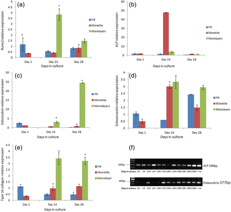
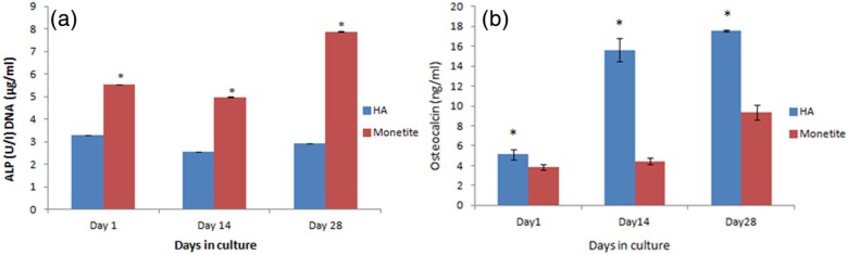
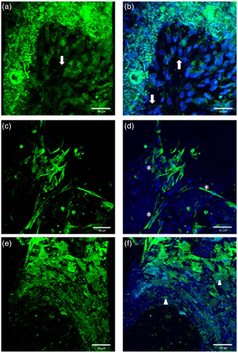
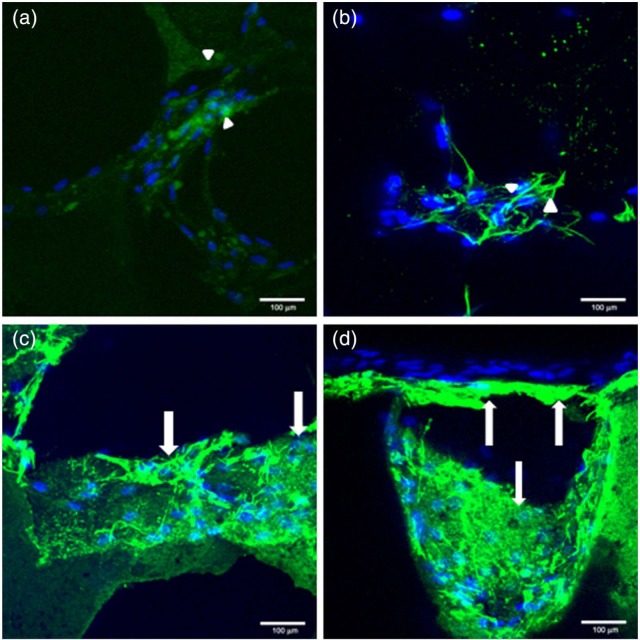
Tissue engineering-based bone grafts are emerging as a viable alternative treatment modality to repair and regenerate tissues damaged as a result of disease or injury. The choice of the biomaterial component is a critical determinant of the success of the graft or scaffold; essentially, it must induce and allow native tissue integration, and most importantly mimic the hierarchical structure of the native bone. Calcium phosphate bioceramics are widely used in orthopaedics and dentistry applications due to their similarity to bone mineral and their ability to induce a favourable biological response. One such material is monetite, which is biocompatible, osteoconductive and has the ability to be resorbed under physiological conditions. The osteoinductive properties of monetite in vivo are known; however, little is known of the direct effect on osteoinduction of human mesenchymal stem cells in vitro. In this study, we evaluated the potential of monetite to induce and sustain human mesenchymal stem cells towards osteogenic differentiation. Human mesenchymal stem cells were seeded on the monetite scaffold in the absence of differentiating factors for up to 28 days. The gene expression profile of bone-specific markers in cells on monetite scaffold was compared to the control material hydroxyapatite. At day 14, we observed a marked increase in alkaline phosphatase, osteocalcin and osteonectin expressions. This study provides evidence of a suitable material that has potential properties to be used as a tissue engineering scaffold.

Keywords: Mesenchymal stem cells; monetite; osteoinduction.


Conflict of interest statement

Declaration of conflicting interests: The authors declare that there is no conflict of interest.

Figures

Figure 1.
Figure 1.
Optical image of monetite produced from brushite scaffold by autoclaving at 121°C for 30 min.
Figure 2.
Figure 2.
X-ray pattern of brushite cement. The green line represents the main brushite peaks (JCPDS file 00-009-0077) and the red line represents the main (Δ) monetite peaks (JCPDS file 00-009-0080). (⋄) Main β-tricalcium phosphate peaks (JCPDS file 00-009-0169).
Figure 3.
Figure 3.
X-ray pattern of monetite scaffold. The red line represents the main monetite peaks (JCPDS file 00-009-0080) and the blue line represents the main (⋄) β-tricalcium phosphate peaks (JCPDS file 00-009-0169).
Figure 4.
Figure 4.
FT-IR spectra of the cements indicating the formation of brushite and monetite. FT-IR: Fourier transform–infrared spectroscopy.
Figure 5.
Figure 5.
The microstructure of (a) HA sample shows rounded crystals and (b) monetite shows needle-like crystals typical of a monetite phase. HA: hydroxyapatite.
Figure 6.
Figure 6.
(a) Runx2 gene expression over 28 days of monetite and HA scaffolds with monolayer cultures seeded with hMSC in non-osteogenic-conditioned medium (p < 0.05), (b) ALP gene expression over 28 days of monetite and HA scaffolds with monolayer cultures seeded with hMSC in non-osteogenic-conditioned medium (p < 0.05), (c) osteocalcin gene expression over 28 days of monetite and HA scaffolds with monolayer cultures seeded with hMSC in non-osteogenic-conditioned medium (p < 0.05), (d) osteonectin gene expression over 28 days of monetite and HA scaffolds with monolayer cultures seeded with hMSC in non-osteogenic-conditioned medium (p < 0.05), (e) type IA collagen gene expression over 28 days of monetite and HA scaffolds with monolayer cultures seeded with hMSC in non-osteogenic-conditioned medium (p < 0.05) and (f) semi-quantitative RT-PCR analysis of HA and monetite scaffolds seeded with hMSCs in non-osteogenic and osteogenic-conditioned medium (M = monetite in non-osteogenic medium, H = HA in non-osteogenic-conditioned medium, M+ = monetite in osteogenic-conditioned medium, H+ = HA in osteogenic-conditioned medium). HA: hydroxyapatite; hMSCs: human mesenchymal stem cells; RT-PCR: reverse transcription–polymerase chain reaction.
Figure 7.
Figure 7.
(a) ALP/DNA content of HA and monetite scaffolds seeded with hMSCs in non-osteogenic-conditioned medium 28 days (p < 0.001) and (b) osteocalcin production of hMSC seeded in HA and monetite in non-osteogenic-conditioned medium (p < 0.01). HA: hydroxyapatite; hMSCs: human mesenchymal stem cells.
Figure 8.
Figure 8.
Confocal laser scanning microscopy (CLSM) – direct staining of actin microfilament cytoskeletal protein and nuclei counterstained with DAPI at day 3 of monolayer culture. DAPI: 4′,6-diamidino-2-phenylindole.
Figure 9.
Figure 9.
CLSM demonstrating the morphology of hMSCs seeded on monetite in non-osteogenic-conditioned media at day 14 (a, c, e) without nuclei staining and (b, d, f) with nuclei staining. (a, b) Direct staining for actin microfilament cytoskeletal proteins, arrows demonstrating the stress fibres for F-actin microfilaments; (c, d) indirect staining for intermediate filaments cytoskeletal protein, asterisks (*) denoting the fibroblast-like cell morphology; and (e, f) indirect staining for adhesion molecule vinculin, arrow heads showing the focal adhesion proteins either within the cell cytoplasm or staining on the scaffold. CLSM: confocal laser scanning microscopy; hMSCs: human mesenchymal stem cells.
Figure 10.
Figure 10.
CLSM demonstrating collagen matrices deposited in non-osteogenic-conditioned medium at day 42 in hMSC seeded in HA and monetite scaffolds counterstained with nuclei (a, b) HA, arrow heads demonstrating low-level staining of type IA collagen, and (c, d) monetite scaffold, arrows demonstrating strong staining surrounding cell nuclei. CLSM: confocal laser scanning microscopy; hMSCs: human mesenchymal stem cells; HA: hydroxyapatite.

References

    1. Bose S, Tarafder S. Calcium phosphate ceramic systems in growth factor and drug delivery for bone tissue engineering: a review. Acta Biomater 2012; 8: 1401–1421. - PMC - PubMed
    1. William DF. On the mechanisms of biocompatibility. Biomaterials 2008; 29: 2941–2953. - PubMed
    1. Olszta MJ, Cheng X, Jee S, et al. Bone structure and formation: a new perspective. Mater Sci Eng R Rep 2007; 58: 77–116.
    1. Hubbell JA. Biomaterials in tissue engineering. Nat Biotechnol 1995; 13: 565–576. - PubMed
    1. Hollister SJ. Porous scaffold design for tissue engineering. Nat Mater 2005; 4: 518–524. - PubMed

LinkOut - more resources