

doi: 10.1016/j.eats.2013.11.007.
eCollection 2014 Apr.
Ultrasound-assisted hip arthroscopy
Affiliations
- PMID: 24904772
- PMCID: PMC4044512
- DOI: 10.1016/j.eats.2013.11.007
Item in Clipboard
Ultrasound-assisted hip arthroscopy
Arthrosc Tech.
.
Abstract
We describe the use of intraoperative ultrasound for the safe development of arthroscopic portals during hip arthroscopy without the requirement for fluoroscopy. We find this technique consistently accurate, allowing the safe introduction of arthroscopic instruments into the hip with a very low rate of iatrogenic injury. We have further developed the technique for application to both central- and peripheral-compartment procedures. We now have a total experience of more than 700 procedures to date. With the described technique of ultrasound guidance for portal placement, fluoroscopy is required in fewer than 2% of hip arthroscopy procedures at our institution.
Figures
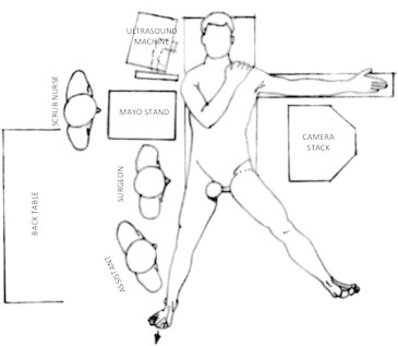
Basic theater layout.
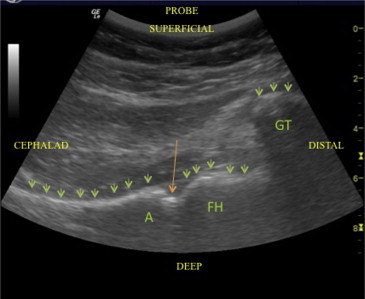
Ultrasound image of right hip (longitudinal view, no traction). The green arrows indicate the bony artifact, and the orange arrow indicates the joint space. (A, acetabulum; FH, femoral head; GT, greater trochanter.)
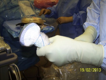
Sterile draping of ultrasound transducer.
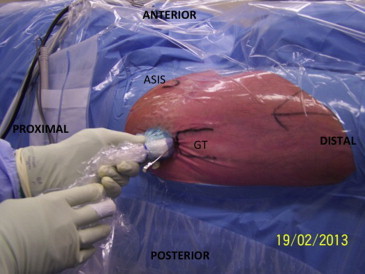
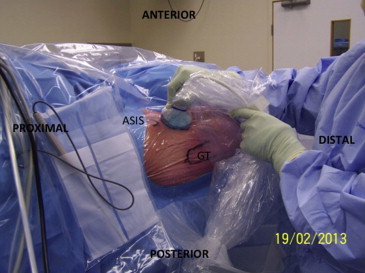
Position of transducer for initial (viewing) portal for central-compartment hip arthroscopy. (ASIS, anterior superior iliac spine; GT, greater trochanter.)
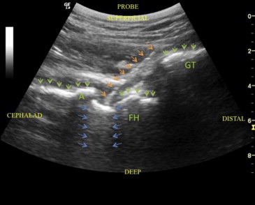
Ultrasound image showing air–intra-articular artifact (light-saber sign) and needle position for initial (viewing) portal placement. The green arrows indicate the bony artifact, the orange arrows indicate the needle, and the blue arrows indicate the light-saber artifact. (A, acetabulum; FH, femoral head; GT, greater trochanter.)
Transducer position for peripheral-compartment initial portal placement—longitudinal view along femoral neck. (ASIS, anterior superior iliac spine; GT, greater trochanter.)
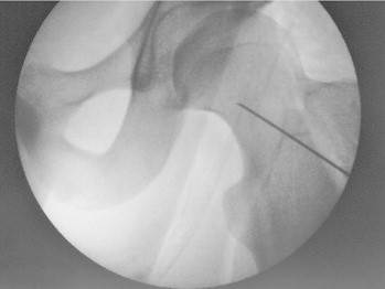
Fluroscopic image demonstrating location of initial (camera) portal placement for peripheral compartment procedures. We now rarely use fluroscopy for peripheral compartment procedures at our institution.
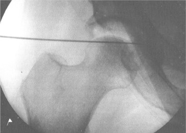
Fluroscopic image of posterior trochanteric central compartment (camera) portal, originally placed under ultrasound guidance. A more posterior position behind the femoral head is easy to obtain using ultrasound which may assist in treating superior acetabular cartilage lesions. We now rarely use fluroscopy for central compartment procedures at our institution.
References
LinkOut - more resources
Full Text Sources
Other Literature Sources
